. 2010 Nov 5;7(5):581-92.
doi: 10.1016/j.stem.2010.10.001.

The steroid hormone ecdysone functions with intrinsic chromatin remodeling factors to control female germline stem cells in Drosophila

Affiliations

The steroid hormone ecdysone functions with intrinsic chromatin remodeling factors to control female germline stem cells in Drosophila

Elizabeth T Ables et al. Cell Stem Cell. .

Abstract

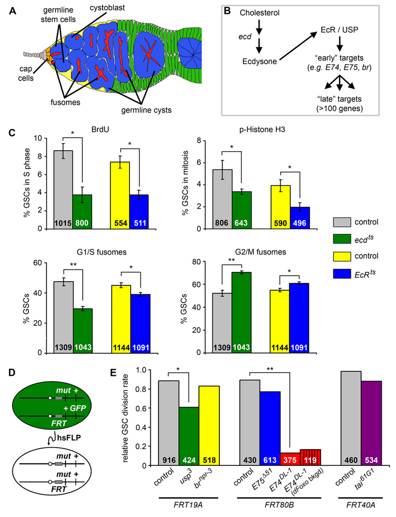
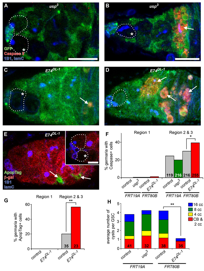
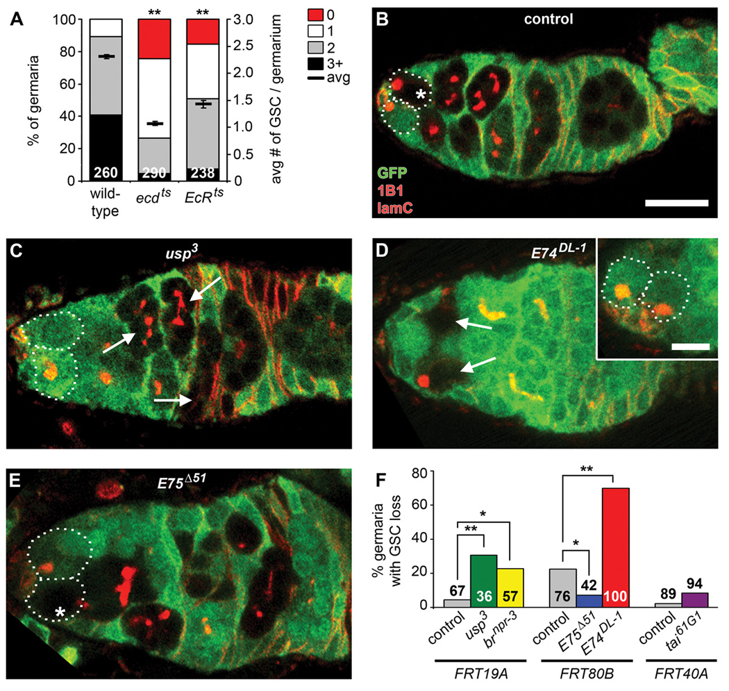
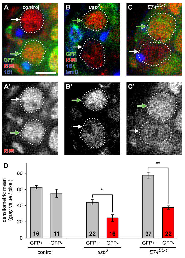
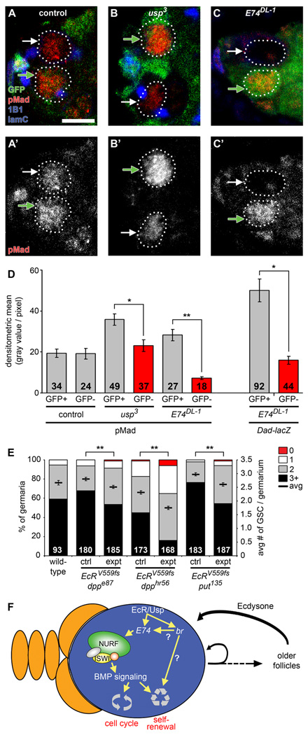
Steroid hormones are known systemic regulators of multiple normal and cancerous tissues; however, whether or how they impact the fate and function of adult stem cells is unclear. In the Drosophila ovary, insulin signals modulate the proliferation and self-renewal of germline stem cells (GSCs), yet despite evidence that additional systemic factors control GSC activity, these have remained largely unknown. Here, we report that ecdysone, a steroid hormone structurally related to mammalian sex steroids, directly regulates adult GSC proliferation and self-renewal independently of insulin signaling. Ecdysone controls GSCs through a functional interaction with the chromatin remodeling factors ISWI, an intrinsic epigenetic factor required for GSC fate and activity, and Nurf301, the largest subunit of the ISWI-containing NURF chromatin remodeling complex. Our findings support a link between systemic steroid hormones and the intrinsic chromatin remodeling machinery as a potential mechanism to promote broad transcriptional programs required for adult stem cell self-renewal.


Conflict of interest statement

The authors declare no competing conflicts of interest.

Figures

Figure 1
Figure 1. Ecdysone acts directly on GSCs to control GSC proliferation at the level of the G2 cell cycle phase
(A) Drosophila germarium. GSCs are identifiable by their typical fusome (red) and attachment to the niche (cap cells, orange; terminal filament cells, gray), and associate with a subset of inner germarial sheath cells (yellow). GSC division produces one self-renewing daughter and one cystoblast that divides to form a 16-cell germline cyst encapsulated by somatic follicle cells (green). (B) Diagram of ecdysone pathway showing components relevant to this study. (C) Frequencies of GSCs in S phase (measured by BrdU incorporation), mitosis (measured by phospho-histone H3 labeling, p-Histone H3), or displaying G1/S or G2/M fusome morphologies in temperature-sensitive ecd1/Df(3L)R-G7 (ecdts) and EcRM554fs/EcRA483T (EcRts) mutants versus heterozygous controls at 29°C for five days. The number of GSCs analyzed is shown inside bars. Error bars, mean ± s.e.m. (D) FLP/FRT technique used to generate genetic mosaics. Mitotic recombination is mediated by heat shock-induced expression of flipase (hsFLP). Homozygous mutant (mut) GSCs are identified by the absence of a GFP marker, which is linked to the wild-type allele. (E) Relative GSC division rates (normalized ratio between GFP-negative and -positive progeny) in wild-type control, usp3, brnpr-3, E75Δ51, E74DL-1, and tai61G1 mosaic germaria. E74DL-1 mosaic germaria were also analyzed in the dFOXO21/dFOXO25 mutant background (dFoxo bkgd). The number of germline cystoblasts/cysts analyzed is shown inside bars. *p < 0.005, **p < 0.001. (See also Figure S1 and Table S2.)
Figure 2
Figure 2. E74DL-1, but not usp3, mosaic germaria display increased cell death of germline cysts in posterior regions of germarium
(A–D) usp3 (A and B) and E74DL-1 (C and D) mosaic germaria labeled with GFP (green), cleaved Caspase 3 (red, apoptotic cells), 1B1 (blue, fusome) and laminC (blue, nuclear envelope of cap cells) at six days after heat shock (AHS). GSCs (outlined) are identified by their fusome morphology and attachment to cap cells. (E) E74DL-1 mosaic germaria labeled with ApopTag (green, apoptotic cells), β-galactosidase (red), 1B1 (blue) and laminC (blue) at six days AHS. In (A–E), mutant clones are recognized by the absence of GFP (A–D) or β-galactosidase (E) expression in GSCs (asterisks). Arrows indicate apoptotic cells. Scale bar represents 10 µm (A–E) or 5 µm (inset in E, showing GSCs from same germarium in different focal plane). (F and G) Percentages of control, usp3, and E74DL-1 mosaic germaria containing cleaved Caspase 3-positive (F) or ApopTag-positive (G) cells in either region 1 (anterior-most region, containing GSCs, cystoblasts, and dividing cysts) or region 2 and 3 (containing 16-cell cysts and associated follicle cells). Number of germaria (F) or number of germaria with a β-galactosidase-negative clone (G) is shown inside bars. *p < 0.05, **p < 0.005. (H) Distribution of the average number of control (GFP-positive) or mutant (GFP-negative) cystoblasts/2-cell cysts, 4-, 8-, and 16-cell cysts (CB & 2 cc, 4 cc, 8 cc, and 16 cc, respectively) per corresponding GSC in E74DL-1 or usp3 mosaic germaria. The number of germaria analyzed is shown inside bars. **p < 0.001 for 4-cell, 8-cell, and 16-cell cysts.
Figure 3
Figure 3. Ecdysone directly controls GSC maintenance predominantly via E74
(A) Frequencies of germaria containing zero, one, two, or three or more GSCs (left Y-axis), and average number of GSCs per germarium (right Y-axis) in ecdts and EcRts mutant versus wild-type females one week after incubation at the restrictive temperature. See Table S1 for heterozygous controls. The number of germaria analyzed is shown inside bars. Error bars, mean ± s.e.m. (B–E) Control, usp3, E74DL-1, and E75Δ51 mosaic germaria labeled with GFP (green), 1B1 (red, fusome) and laminC (red, nuclear envelope of cap cells) at 10 days AHS (outlines indicate GSCs). Control and mutant clones are recognized by the absence of GFP expression in GSCs (asterisks) and their progeny (B and E) or just in their progeny (arrows), if the GSC has been lost from the niche (C and D). Scale bar represents 10 µm (B–E) or 5 µm (inset in D, showing GSCs from same germarium in different focal plane). (F) Frequencies of germaria with control, usp3, brnpr-3, E75Δ51, E74DL-1, and tai61G1 mosaic germline showing GSC loss at 10 days AHS. Number of germaria with mosaic germline analyzed is shown inside or above bars. *p < 0.05, **p < 0.001. (See also Figure S1 and Tables S1 and S2.)
Figure 4
Figure 4. Ecdysone signaling affects nuclear ISWI protein levels
(A–C) Control (A), usp3 (B), and E74DL-1 (C) mosaic germaria analyzed six days AHS and labeled with GFP (green), ISWI (red), 1B1 (blue, fusomes) and laminC (in B, blue, nuclear envelope of cap cells). Control and mutant GSCs (outlined) are recognized by the absence of GFP expression (GFP-negative GSCs, white arrows), while cells that did not undergo mitotic recombination are GFP-positive (GFP-positive GSCs, green arrows). Scale bar represents 5 µm. (D) Quantification of nuclear ISWI protein levels in control and mutant GSCs. The number of GSCs analyzed is shown inside bars. Error bars, mean ± s.e.m. *p < 0.05, **p < 0.001. (See also Figure S3.)
Figure 5
Figure 5. Ecdysone signaling functionally interacts with iswi and nurf301 to control the maintenance of GSCs
(A and B) Frequencies of germaria containing zero, one, two, or three or more GSCs (left Y-axis), and average number of GSCs per germarium (right Y-axis) in seven-day old wild-type females versus single heterozygotes (ctrl) or double heterozygotes (expt) for mutations in iswi, nurf301, EcR, and InR (A), and iswi, E75, E74, and br (B). The same wild-type data are shown in (A) and (B). Additional results are shown in Table 1. The number of germaria analyzed is shown inside bars. Error bars, mean ± s.e.m. **p < 0.001. (See also Figure S2.)
Figure 6
Figure 6. The ecdysone pathway affects BMP signaling levels in GSCs and genetically interacts with dpp and put to control GSC maintenance
(A–C) Control (A), usp3 (B), and E74DL-1 (C) mosaic germaria analyzed six days AHS and labeled with GFP (green), pMad (red), 1B1 (blue, fusomes) and laminC (blue, nuclear envelope of cap cells). Control and mutant GSCs (outlined) are recognized by the absence of GFP expression (GFP-negative GSCs indicated by white arrows), while cells that did not undergo mitotic recombination are GFP-positive (GFP-positive GSCs indicated by green arrows). Scale bar represents 5 µm. (D) Quantification of nuclear pMad protein levels in control, usp3 and E74DL-1 GSCs (left) and nuclear Dad-lacZ levels in E74DL-1 GSCs (right). The number of GSCs analyzed is shown inside bars. (E) Frequencies of germaria containing zero, one, two, or three or more GSCs (left Y-axis), and average number of GSCs per germarium (right Y-axis) in seven-day old wild-type females versus single heterozygotes (ctrl) or double heterozygotes (expt) for mutations in EcR, dpp, and put. Wild-type data are the same shown in Figure 5. Additional results are shown in Table 1. The number of germaria analyzed is shown inside bars. Error bars, mean ± s.e.m. *p < 0.005, **p < 0.001. (F) Proposed model for how a GSC (blue) within its niche (orange) is controlled by an interaction between the steroid hormone ecdysone and the intrinsic epigenetic machinery (see text for details).

Similar articles

Cited by

References

    1. Asselin-Labat ML, Vaillant F, Sheridan JM, Pal B, Wu D, Simpson ER, Yasuda H, Smyth GK, Martin TJ, Lindeman GJ, et al. Control of mammary stem cell function by steroid hormone signalling. Nature. 2010;465:798–802. - PubMed
    1. Badenhorst P, Voas M, Rebay I, Wu C. Biological functions of the ISWI chromatin remodeling complex NURF. Genes & Dev. 2002;16:3186–3198. - PMC - PubMed
    1. Badenhorst P, Xiao H, Cherbas L, Kwon SY, Voas M, Rebay I, Cherbas P, Wu C. The Drosophila nucleosome remodeling factor NURF is required for Ecdysteroid signaling and metamorphosis. Genes & Dev. 2005;19:2540–2545. - PMC - PubMed
    1. Bai J, Uehara Y, Montell DJ. Regulation of invasive cell behavior by taiman, a Drosophila protein related to AIB1, a steroid receptor coactivator amplified in breast cancer. Cell. 2000;103:1047–1058. - PubMed
    1. Bialecki M, Shilton A, Fichtenberg C, Segraves WA, Thummel CS. Loss of the ecdysteroid-inducible E75A orphan nuclear receptor uncouples molting from metamorphosis in Drosophila. Dev Cell. 2002;3:209–220. - PubMed

Publication types