. 2011 Feb;32(5):1291-300.
doi: 10.1016/j.biomaterials.2010.10.018. Epub 2010 Oct 30.

Hierarchical and non-hierarchical mineralisation of collagen

Affiliations

Hierarchical and non-hierarchical mineralisation of collagen

Yan Liu et al. Biomaterials. 2011 Feb.

Abstract

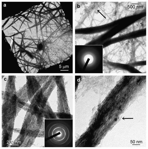
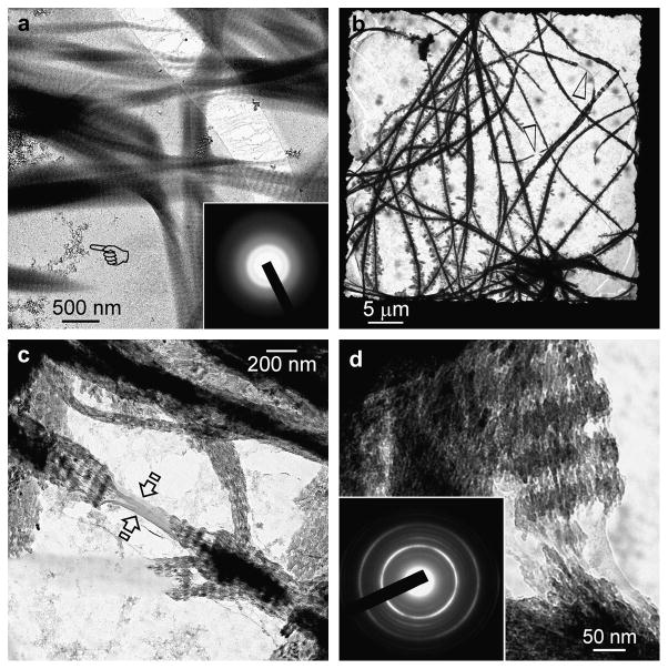
Biomineralisation of collagen involves functional motifs incorporated in extracellular matrix protein molecules to accomplish the objectives of stabilising amorphous calcium phosphate into nanoprecursors and directing the nucleation and growth of apatite within collagen fibrils. Here we report the use of small inorganic polyphosphate molecules to template hierarchical intrafibrillar apatite assembly in reconstituted collagen in the presence of polyacrylic acid to sequester calcium and phosphate into transient amorphous nanophases. The use of polyphosphate without a sequestration analogue resulted only in randomly-oriented extrafibrillar precipitations along the fibrillar surface. Conversely, the use of polyacrylic acid without a templating analogue resulted only in non-hierarchical intrafibrillar mineralisation with continuous apatite strands instead of discrete crystallites. The ability of using simple non-protein molecules to recapitulate different levels of structural hierarchy in mineralised collagen signifies the ultimate simplicity in Nature's biomineralisation design principles and challenges the need for using more complex recombinant matrix proteins in bioengineering applications.


Figures

Figure 1
Figure 1
Unstained TEM showing only extrafibrillar mineralisation in the templating analogue control (no sequestration analogue in mineralisation medium). (a) After 24 hours, partially-coalesced ACPs (arrow) were attached to the fibril surface (open arrowhead) but were too large to penetrate the collagen fibril. (b) An unmineralised collagen fibril (pointer) with heavier ACP surface aggregation. (c) Some ACP phases were transformed into immature finger-like immature apatite (inset). (d) After 72 hours, spherules of needle-shaped apatite crystallites (inset; > 100 nm long) coated the surface (open arrow) of unmineralised collagen fibrils (open arrowhead).
Figure 2
Figure 2
Unstained TEM showing the temporal events associated with non-hierarchical intrafibrillar mineralisation in the sequestration analogue control (no templating analogue). (a) Grid space showing swollen electron-dense collagen fibrils (asterisk) after 24 hours. (b) Swollen, electron-dense fibrils with a smooth appearance. ACP nanoparticle clusters were seen predominantly around unmineralised fibrils (arrow). ACP nanoparticles appeared to have penetrated the swollen fibrils and coalesced into a continuous amorphous mineral phase (inset). (c) After 72 h of mineralization, collagen fibrils were no longer swollen and exhibited a fibrous appearance. Although they contained intrafibrillar mineral, they lacked cross-banding patterns. Inset: Selected are electron diffraction (SAED) of individual mineralised fibrils produced ring patterns that are characteristic of apatite (note arc-shaped patterns indicating that minerals are arranged along the longitudinal axis of the collagen fibrils – see also Supplementary Fig.4). (d) High magnification of a mineralised fibril showing the continuity of the mineral strands and absence of discrete crystallites (arrow).
Figure 3
Figure 3
Unstained TEM showing the spatial effects of different templating analogue concentrations on the amorphous stage of hierarchical intrafibrillar mineralisation in the presence of a sequestration analogue. Sodium trimetaphosphate (a) 0.313 wt%, (b) 0.625 wt%, (c) 1.25 wt%, (d) 2.5 wt% was used as the example. Only ACP nanoparticle aggregates were identified along the collagen fibril surfaces (open arrowheads). Large ACP phases (see Supplementary Figure 2) were not involved in intrafibrillar mineralisation.
Figure 4
Figure 4
Unstained TEM showing the spatial effects of different templating analogue concentrations on the crystalline stage of hierarchical intrafibrillar mineralisation in the presence of a sequestration analogue. Sodium tripolyphosphate (a) 0.313%, (b) 0.625%, (c) 1.25%, (d) 2.5% was used as the example. The similarity between Fig. 4(a) and Fig. 2(c) suggests that 0.313 wt% sodium tripolyphosphate is insufficient to produce hierarchical intrafibrillar banding. Inset in (c) indicates crystalline phase that is characteristic of apatite and is applicable to all subfigures. Partially-mineralised collagen fibrils were illustrated as the heavily-mineralised fibrils were too electron-dense for cross-banding to be discerned in bulk, unsectioned fibrils.
Figure 5
Figure 5
Unstained TEM of collagen treated with 2.5 wt% sodium tripolyphosphate and mineralised in polyacrylic acid-containing SBF. (A) Swollen, cross-banded collagen fibrils containing amorphous electron-dense minerals (inset) after 24 h. (b) Grid space showing heavy mineralisation of the collagen fibrils after 72 h. A few fibrils contained unmineralised regions (open arrowheads). (c) Partially-mineralised collagen fibril with cross-banding created by intrafibrillar mineral assembly. Absence of minerals in the unmineralised region (between open arrows) resulted in extensive shrinkage during chemical dehydration for TEM. (d) Partially-mineralised collagen fibril showing hierarchical arrangement of overlapping nanoplatelets. Inset: SAED with ring patterns corresponding to those of poorly-crystalline apatite (note: arc-shaped patterns indicating that minerals are arranged along the longitudinal axis of the collagen fibrils – see also Supplementary Fig.4).
Figure 6
Figure 6
Infrared spectra (1800–700 cm−1) of type I collagen before and after treatment with 2.5 wt% sodium tripolyphosphate and after subjected to competitive desorption with 250 mM NaCl for 24 h. After tripolyphosphate treatment, new phosphate peaks could be identified (asterisks). There was also a simultaneous reduction in the series of collagen bands between Amide II and III (arrows). After 24 h of NaCl desorption, the spectrum was similar to that of the original type I collagen. This suggests that the sodium polyphosphate binding mechanism is exclusively electrostatic in nature, probably via the formation of ionic bridges.
Figure 7
Figure 7
Schematic showing the different status of apatite deposition around and within a collagen fibril in the presence of different combinations of sequestration and templating analogues.
Figure 8
Figure 8
Schematic depicting possible ionic cross-linking involved with binding of sodium tripolyphosphate anions to collagen molecules. Free hydroxyl ions that do not interact with collagen molecules contribute to recruitment of polyacrylic acid-stabilised amorphous calcium phosphate nanoprecursors and induction of apatite nucleation.

References

    1. Wong TS, Brough B, Ho CM. Creation of functional micro/nano systems through top-down and bottom-up approaches. Mol Cell Biomech. 2009;6:1–55. - PMC - PubMed
    1. Traub W, Arad T, Weiner S. Three-dimensional ordered distribution of crystals in turkey tendon collagen fibers. Proc Natl Acad Sci USA. 1989;86:9822–9826. - PMC - PubMed
    1. Wang L, Nancollas GH. Pathways to biomineralization and biodemineralization of calcium phosphates: the thermodynamic and kinetic controls. Dalton Trans. 2009;15:2665–2672. - PubMed
    1. Saito T, Arsenault AL, Yamauchi M, Kuboki Y, Crenshaw MA. Mineral induction by immobilized phosphoproteins. Bone. 1997;21:305–311. - PubMed
    1. Cölfen H. Bio-inspired mineralization using hydrophilic polymers. Top Curr Chem (Springer-Verlag, Berlin: Heidelberg) 2007;271:1–77.

Publication types

MeSH terms