

Dual-mode IVUS catheter for intracranial image-guided hyperthermia: feasibility study
- PMID: 21041144
- PMCID: PMC3018697
- DOI: 10.1109/TUFFC.2010.1723
Dual-mode IVUS catheter for intracranial image-guided hyperthermia: feasibility study
Abstract
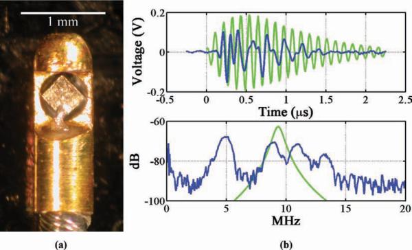
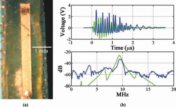
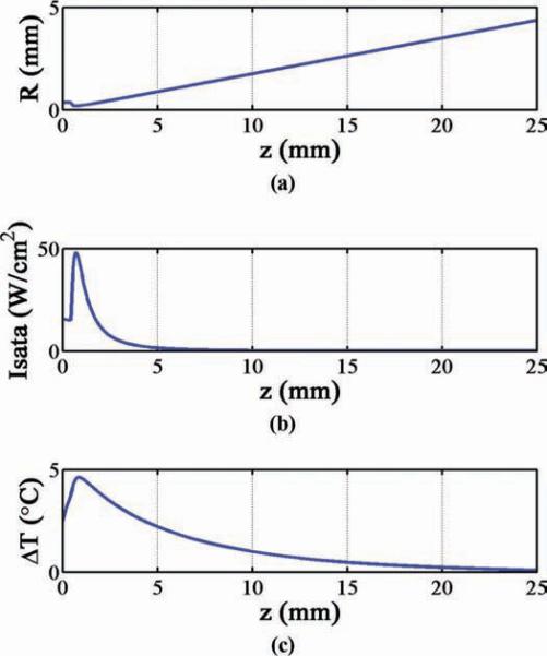
In this study, we investigated the feasibility of modifying 3-Fr IVUS catheters in several designs to potentially achieve minimally-invasive, endovascular access for image-guided ultrasound hyperthermia treatment of tumors in the brain. Using a plane wave approximation, target frequencies of 8.7 and 3.5 MHz were considered optimal for heating at depths (tumor sizes) of 1 and 2.5 cm, respectively. First, a 3.5-Fr IVUS catheter with a 0.7-mm diameter transducer (30 MHz nominal frequency) was driven at 8.6 MHz. Second, for a low-frequency design, a 220-μm-thick, 0.35 x 0.35-mm PZT-4 transducer--driven at width-mode resonance of 3.85 MHz--replaced a 40-MHz element in a 3.5-Fr coronary imaging catheter. Third, a 5 x 0.5-mm PZT-4 transducer was evaluated as the largest aperture geometry possible for a flexible 3-Fr IVUS catheter. Beam plots and on-axis heating profiles were simulated for each aperture, and test transducers were fabricated. The electrical impedance, impulse response, frequency response, maximum intensity, and mechanical index were measured to assess performance. For the 5 x 0.5-mm transducer, this testing also included mechanically scanning and reconstructing an image of a 2.5-cm-diameter cyst phantom as a preliminary measure of imaging potential.
Figures


Similar articles
-
Intracranial dual-mode IVUS and hyperthermia using circular arrays: preliminary experiments.Ultrason Imaging. 2013 Jan;35(1):17-29. doi: 10.1177/0161734612469372. Ultrason Imaging. 2013. PMID: 23287504 Free PMC article.
-
Dual-mode IVUS transducer for image-guided brain therapy: preliminary experiments.Ultrasound Med Biol. 2011 Oct;37(10):1667-76. doi: 10.1016/j.ultrasmedbio.2011.06.017. Epub 2011 Aug 19. Ultrasound Med Biol. 2011. PMID: 21856073 Free PMC article.
-
Dual-mode intracranial catheter integrating 3D ultrasound imaging and hyperthermia for neuro-oncology: feasibility study.Ultrason Imaging. 2009 Apr;31(2):81-100. doi: 10.1177/016173460903100201. Ultrason Imaging. 2009. PMID: 19630251 Free PMC article.
-
Principles and devices.Semin Vasc Surg. 2006 Sep;19(3):128-31. doi: 10.1053/j.semvascsurg.2006.06.006. Semin Vasc Surg. 2006. PMID: 16996413 Review.
-
High-frequency intracoronary ultrasound imaging.Semin Interv Cardiol. 1997 Mar;2(1):33-41. Semin Interv Cardiol. 1997. PMID: 9546982 Review.
Cited by
-
Intracranial dual-mode IVUS and hyperthermia using circular arrays: preliminary experiments.Ultrason Imaging. 2013 Jan;35(1):17-29. doi: 10.1177/0161734612469372. Ultrason Imaging. 2013. PMID: 23287504 Free PMC article.
-
Dual-mode IVUS transducer for image-guided brain therapy: preliminary experiments.Ultrasound Med Biol. 2011 Oct;37(10):1667-76. doi: 10.1016/j.ultrasmedbio.2011.06.017. Epub 2011 Aug 19. Ultrasound Med Biol. 2011. PMID: 21856073 Free PMC article.
-
Impedance matching network for high frequency ultrasonic transducer for cellular applications.Ultrasonics. 2016 Feb;65:258-67. doi: 10.1016/j.ultras.2015.09.016. Epub 2015 Sep 28. Ultrasonics. 2016. PMID: 26442434 Free PMC article.
-
Contrast Enhanced Superharmonic Imaging for Acoustic Angiography Using Reduced Form-Factor Lateral Mode Transmitters for Intravascular and Intracavity Applications.IEEE Trans Ultrason Ferroelectr Freq Control. 2017 Feb;64(2):311-319. doi: 10.1109/TUFFC.2016.2619687. Epub 2016 Oct 20. IEEE Trans Ultrason Ferroelectr Freq Control. 2017. PMID: 27775903 Free PMC article.
-
An IVUS transducer for microbubble therapies.IEEE Trans Ultrason Ferroelectr Freq Control. 2014 Mar;61(3):441-9. doi: 10.1109/TUFFC.2014.2929. IEEE Trans Ultrason Ferroelectr Freq Control. 2014. PMID: 24569249 Free PMC article.
References
-
- Cancer Facts & Figures 2008. American Cancer Society; Atlanta, GA: 2008.
-
- Kleihues P, Cavenee WK. Pathology and Genetics of Tumours of the Nervous System. 2nd ed. IARC Press; Lyon, France: 2000. International Agency for Research on Cancer, International Society of Neuropathology, International Academy of Pathology, and Preuss Foundation for Brain Tumor Research.
-
- Statistical Report: Primary Brain Tumors in the United States, 2000–2004. Central Brain Tumor Registry of the United States; Hinsdale, IL: 2008.
-
- Mareel M. Anti-invasive brain tumor therapy: General aspects and future strategies. In: Mikkelsen T, editor. Brain Tumor Invasion: Biological, Clinical, and Therapeutic Considerations. Wiley-Liss; New York, NY: 1998. p. 393.
-
- Wilkins RH, Rengachary SS. Neurosurgery. 2nd ed. McGraw-Hill; New York, NY: 1996.
