. 2011 Jan 15;20(2):354-67.
doi: 10.1093/hmg/ddq470. Epub 2010 Nov 1.

Endothelial cells from humans and mice with polycystic kidney disease are characterized by polyploidy and chromosome segregation defects through survivin down-regulation

Affiliations

Endothelial cells from humans and mice with polycystic kidney disease are characterized by polyploidy and chromosome segregation defects through survivin down-regulation

Wissam A AbouAlaiwi et al. Hum Mol Genet. .

Abstract

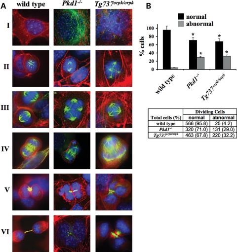
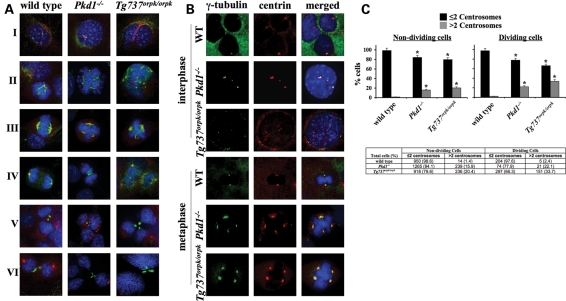
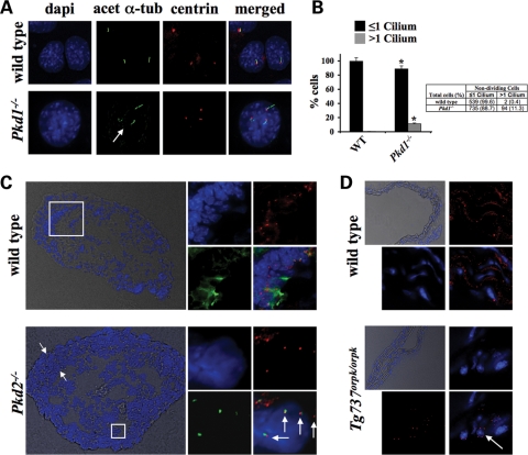
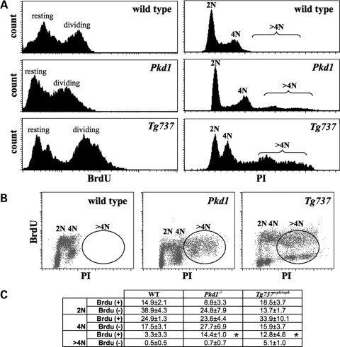
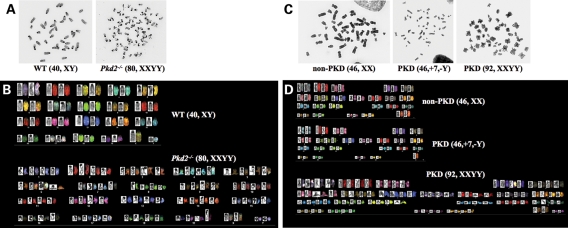
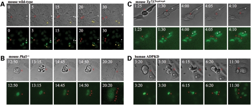
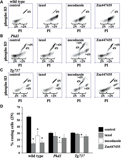
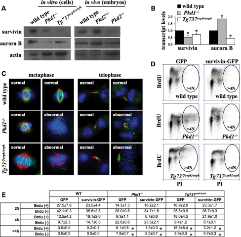
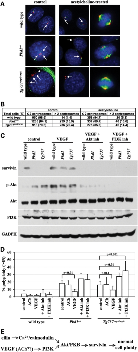
Autosomal-dominant polycystic kidney disease (ADPKD) is the most common hereditary and systemic disorder associated with various cardiovascular complications. It has been implicated with dysfunction in primary cilia. We and others have shown that the immediate function of endothelial cilia is to sense extracellular signal. The long-term function of cilia is hypothesized to regulate cell cycle. Here, we show that ciliary function (polycystins) and structure (polaris) are required for proper cellular division. Cilia mutant cells undergo abnormal cell division with apparent defects in mitotic spindle formation, cellular spindle assembly checkpoint and centrosome amplification. Down-regulation of the chromosomal passenger survivin contributes to these abnormalities, which further result in cell polyploidy. Re-expression of survivin restores a competent spindle assembly checkpoint and reduces polyploidy. Aged animals show a more severe phenotype in cellular division, consistent with progression of cardiovascular complications seen in older ADPKD patients. For the first time, we show that structure and function of mechanosensory cilia are crucial in maintaining proper cellular proliferation. Furthermore, developmental aging plays a crucial role in the progression of these abnormal cellular phenotypes. We propose that abnormal function or structure of primary cilia not only causes failure to transmit extracellular signals, but also is associated with cytokinesis defects in both mice and humans with polycystic kidney disease.


Figures

Figure 1.
Figure 1.
Cell division in cilia mutant cells is characterized by mitotic abnormalities and multipolar spindle formation. (A) Endothelial wild-type, Pkd1−/− and Tg737Orpk/Orpk cells were immunostained with DAPI (blue), acetylated-α-tubulin (green; acet-α-tubulin) and phalloidin (red; actin) to visualize nucleus, cilium, mitotic spindle and actin cytoskeleton. Images were captured at different cell-cycle stages of interphase (I), prophase (II), metaphase (III), anaphase (IV), telophase (V) and cytokinesis (VI). (B) Abnormal dividing cells, indicated as percentages (bar graph) and number of dividing cells (table), are significantly greater in cells with abnormal cilia function (Pkd1−/−) and structure (Tg737Orpk/Orpk) than in wild-type cells. Original magnification, ×100. Asterisks denote significant difference toward corresponding wild-type groups.
Figure 2.
Figure 2.
Cilia mutant cells are characterized by centrosome overduplication and abnormal cell division. (A) Endothelial wild-type, Pkd1−/− and Tg737Orpk/Orpk cells were immunostained with DAPI (blue), acetylated-α-tubulin (green; acet-α-tubulin) and pericentrin (red; centrin) to visualize nucleus, cilium, mitotic spindle and centrosome. Images were captured at different cell-cycle stages of interphase (I), prophase (II), metaphase (III), anaphase (IV), telophase (V) and cytokinesis (VI). (B) To confirm the specificity of centrosome localization, immunostaining was carried out with γ-tubulin (green) and pericentrin (red) in wild-type, Pkd1−/− and Tg737Orpk/Orpk non-dividing (interphase) and dividing (metaphase) cells. (C) Compared with wild-type cells, Pkd1−/− and Tg737Orpk/Orpk cells have significantly more centrosomes in both dividing and non-dividing stages. The number of dividing and non-dividing cells containing ≤2 or >2 centrosomes is indicated as percentages (bar graph) or total cell counts (table). Original magnification, ×100. Asterisks denote significant difference toward corresponding wild-type groups.
Figure 3.
Figure 3.
Centrosome overduplication results in multiple cilia formation in vitro and in vivo. (A) Endothelial wild-type (WT) and Pkd1−/− cells were immunostained with DAPI (blue), acetylated-α-tubulin (green; acet-α-tubulin) and pericentrin (red; centrin) to visualize nucleus, cilium and centrosome. (B) Compared with wild-type cells, Pkd1−/− cells have more cilia. Scores of randomly chosen non-dividing cells containing one or more cilia are indicated as percentages (bar graph) or total cell counts (table). (C) Aorta sections from wild-type and Pkd2−/− 15.5-day embryos were stained with acetylated α-tubulin (green), pericentrin (red) and counterstained with DAPI (blue) to visualize cilia, centrosome and nucleus, respectively. The boxes indicate areas of arteries that were further magnified, as shown in the insets, which reveal individual cells with multiple cilia and centrosomes in Pkd2−/−, but not in wild-type, aortas. Arrows indicate thickening of intima tissues. (D) Aorta sections from wild-type and Tg737Orpk/Orpk adult mice were stained with pericentrin (red) and counterstained with DAPI. The boxes indicate areas of arteries that were further magnified, as shown in the insets, which reveal individual cells with multiple centrosomes in Tg737Orpk/Orpk, but not in wild-type, aortas. Original magnification, ×100, except for those in phase contrast, ×20. Asterisks denote significant difference toward corresponding wild-type groups. Arrows indicate multiple primary cilia and/or centrosomes.
Figure 4.
Figure 4.
Cells with abnormal cilia structure or function are characterized by cell polyploidy. (A) Endothelial wild-type, Pkd1−/− and Tg737Orpk/Orpk cells were analyzed by flow cytometry by PI and BrdU labeling. Representative BrdU and PI labeling profiles present an apparent polyploidy in Pkd1−/− and Tg737Orpk/Orpk cells, but not in wild-type cells. (B) Further analysis indicates that polyploidy Pkd1−/− and Tg737Orpk/Orpk cells are able to undergo mitosis. (C) Evaluation of the percentage of cells with normal DNA content (2N and 4N) shows that Pkd1−/− and Tg737Orpk/Orpk cells contain a greater DNA content (>4N), compared with wild-type cells. The number of dividing and non-dividing cells containing 2N, 4N and >4N DNA content is presented as percentages in the table. Asterisks denote significant difference toward corresponding wild-type groups. N > 5 for each genotype in flow cytometry experiments.
Figure 5.
Figure 5.
Cilia mutant cells are characterized by genomic instability. Chromosomal identifications were carried out in mouse and human cells with abnormal function/structure. (A) Freshly isolated endothelial cells from wild-type and Pkd2−/− embryonic E15.5 aortas were studied for their genomic compositions. A simple chromosome count indicates the presence of polyploidy cells in Pkd2−/−. (B) Further characterization of individual chromosomes was performed with fluorescence probes. Identification of individual chromosomes indicates the tetraploidy nature of Pkd2−/− cells, suggesting abnormal segregation of chromosomes in cilia mutant cells. (C) A single endothelium from interlobar arteries of an ADPKD patient was examined for a simple chromosome count. (D) Further characterization of individual chromosomes was performed with fluorescence probes. Genomic instability is further characterized by addition or reduction on a specific chromosome. Abnormal segregation of chromosomes is also apparent in cells from patients with ADPKD.
Figure 6.
Figure 6.
Mouse and human cilia mutant cells are characterized by the loss of mitotic spindle checkpoint. Live-imaging studies were performed to identify the cause of genomic stability in isolated endothelial cells from wild-type (A), Pkd1−/− (B) and Tg737Orpk/Orpk (C) mice. Isolated primary endothelial cells from a patient with ADPKD were also analyzed. (D) In wild-type endothelial cells, normal cell division results in two daughter cells through an even, bipolar chromosomal segregation. In mouse Pkd or primary human ADPKD cells, the DNA was condensed and was able to line up for chromosomal segregations. During anaphase, however, the cells were not able to separate the chromosomes equally. As a result, cytokinesis did not take place, and cells became polyploidy. The movies show that cilia mutant cells could complete the mitotic checkpoint assembly, but failed to maintain spindle tension during anaphase. Numbers indicate time in minutes and seconds as illustrated in the Supplementary Material, Movies S1–S4. Top panels are phase-contrast images to identify numbers of cells; bottom panels are fluorescence images of Hoechst. Arrows indicate cell boundary in phase contrast or cell nucleus in fluorescence images; each color represents one parental cell. Original magnification, ×40.
Figure 7.
Figure 7.
Cilia mutant cells are characterized by aberrant chromosomal passenger protein. Mitotic-stress tests were performed with taxol to stabilize spindle tubules, nocodazole to depolymerize spindle tubules or Zm447435 to inhibit the function of chromosomal passenger protein. Vehicle treatments are indicated as control. Phosphorylated histone 3b (phospho H3) is used as a marker for nuclear division, and PI is to indicate DNA content. In wild-type cells (A), treatments with taxol, nocodazole or Zm447435 resulted in mitotic arrest. In Pkd1−/− (B) and Tg737Orpk/Orpk (C) cells, only nocodazole promoted mitotic arrest when compared with their corresponding controls. Consistent with the results, further inhibition of chromosomal passenger protein with Zm447435 could not promote mitotic arrest and that chromosomal passenger protein was required for mitotic arrest by taxol. Mitotic-stress test was measured by analyzing changes in resting cells (2N), which are PI and phospho-histone-negative, as indicated by the brackets. (D) These changes are reflected in % non-arrested cells from the total cell population. Asterisks denote significant difference toward corresponding control groups.
Figure 8.
Figure 8.
Survivin plays an important role in genomic instability of cilia mutant cells in vitro and in vivo. (A) Survivin expression was down-regulated in Pkd1−/− and Tg737Orpk/Orpk cells compared with wild-type cells. Likewise, survivin was down-regulated in Pkd2−/− embryos compared with wild-type embryos. Actin expression was used as a loading control. (B) Survivin transcripts were repressed in both Pkd1−/− and Tg737Orpk/Orpk cells. (C) Counterstaining with acetylated-α-tubulin (red) and DAPI (blue) was used to examine subcellular localization of survivin-GFP (green)-transfected cells at metaphase and telophase. (D) Flow cytometry studies show a decrease in polyploidy index in cilia mutant cells transfected with survivin-GFP, but not in those transfected with vector-GFP (empty vector, GFP). Because of the low transfection efficiency in the cells, only transfected (GFP-positive) cells were analyzed. Representative BrdU and PI labeling profiles show an apparent polyploidy in vector-only transfected Pkd1−/− and Tg737Orpk/Orpk cells. Polyploidy Pkd1−/− and Tg737Orpk/Orpk cells are substantially lower in those transfected with survivin-GFP. (E) The number of dividing and non-dividing cells containing 2N, 4N and >4N DNA content is presented as percentages in the table. Asterisks denote significant difference toward corresponding wild-type groups and significant difference between corresponding GFP and survivin-GFP groups.
Figure 9.
Figure 9.
Effect of VEGF on cell ploidy is reversible by PI3K and Akt/PKB inhibitors. (A) Cells with and without 10 μm acetylcholine (ACh) treatment were analyzed with acetylated α-tubulin (green), pericentrin (red) and DAPI (blue) during resting or dividing state. Arrows indicate centrosomes. (B) Their centrosome numbers were calculated and presented in the table. Cells were treated with or without 40 ng/ml of VEGF, 10 μm Akt/PKB inhibitor (Akt inh) or 20 μm PI3K inhibitor (PI3K inh). (C) The cells were analyzed for survivin, phosphorylated-Akt (p-Akt), Akt, PI3K and GADPH. (D) Polyploid cells were studied with flow cytometry and plotted in the graph. (E) A hypothetical pathway is depicted to assess the molecular mechanism of polyploidy.
Figure 10.
Figure 10.
Aging compounds the severity of cilia mutant on centrosome number and cell polyploidy. Aortic endothelial cells were freshly isolated from E15.5-day embryos, 3-week-old pup and 3-month-old adult of wild-type or Tg737Orpk/Orpk mice (C3H background). (A) Cells were stained with pericentrin (red), acetylated-α-tubulin (green) and DAPI (blue) to count the centrosome numbers of resting and dividing cells. Arrows indicate centrosomes. (B) Percentage of abnormal centrosome number from three to four isolations was averaged and plotted. (C) Flow cytometry was also used to analyze cell polyploidy for each age group. (D) Percentage of polyploidy cells from three to four isolations was averaged and plotted. Asterisks denote significant difference toward corresponding wild-type groups.

References

    1. Harris P.C., Torres V.E. Polycystic kidney disease. Annual Rev. Med. 2009;60:321–337. doi:10.1146/annurev.med.60.101707.125712. - DOI - PMC - PubMed
    1. AbouAlaiwi W.A., Takahashi M., Mell B.R., Jones T.J., Ratnam S., Kolb R.J., Nauli S.M. Ciliary polycystin-2 is a mechanosensitive calcium channel involved in nitric oxide signaling cascades. Circ. Res. 2009;104:860–869. doi:10.1161/CIRCRESAHA.108.192765. - DOI - PMC - PubMed
    1. Nauli S.M., Kawanabe Y., Kaminski J.J., Pearce W.J., Ingber D.E., Zhou J. Endothelial cilia are fluid shear sensors that regulate calcium signaling and nitric oxide production through polycystin-1. Circulation. 2008;117:1161–1171. doi:10.1161/CIRCULATIONAHA.107.710111. - DOI - PMC - PubMed
    1. Poelmann R.E., Van der Heiden K., Gittenberger-de Groot A., Hierck B.P. Deciphering the endothelial shear stress sensor. Circulation. 2008;117:1124–1126. doi:10.1161/CIRCULATIONAHA.107.753889. - DOI - PubMed
    1. Nauli S.M., Alenghat F.J., Luo Y., Williams E., Vassilev P., Li X., Elia A.E., Lu W., Brown E.M., Quinn S.J., et al. Polycystins 1 and 2 mediate mechanosensation in the primary cilium of kidney cells. Nat. Genet. 2003;33:129–137. doi:10.1038/ng1076. - DOI - PubMed

Publication types

MeSH terms

Substances