. 2010 Dec 31;285(53):41541-8.
doi: 10.1074/jbc.M110.170894. Epub 2010 Nov 1.

GRK5 deficiency accelerates {beta}-amyloid accumulation in Tg2576 mice via impaired cholinergic activity

Affiliations

GRK5 deficiency accelerates {beta}-amyloid accumulation in Tg2576 mice via impaired cholinergic activity

Shaowu Cheng et al. J Biol Chem. .

Abstract

Membrane G protein-coupled receptor kinase 5 (GRK5) deficiency is linked to Alzheimer disease, yet its precise roles in the disease pathogenesis remain to be delineated. We have previously demonstrated that GRK5 deficiency selectively impairs desensitization of presynaptic M2 autoreceptors, which causes presynaptic M2 hyperactivity and inhibits acetylcholine release. Here we report that inactivation of one copy of Grk5 gene in transgenic mice overexpressing β-amyloid precursor protein (APP) carrying Swedish mutations (Tg2576 or APPsw) resulted in significantly increased β-amyloid (Aβ) accumulation, including increased Aβ(+) plaque burdens and soluble Aβ in brain lysates and interstitial fluid (ISF). In addition, secreted β-APP fragment (sAPPβ) also increased, whereas full-length APP level did not change, suggesting an alteration in favor of β-amyloidogenic APP processing in these animals. Reversely, perfusion of methoctramine, a selective M2 antagonist, fully corrected the difference between the control and GRK5-deficient APPsw mice for ISF Aβ. In contrast, a cholinesterase inhibitor, eserine, although significantly decreasing the ISF Aβ in both control and GRK5-deficient APPsw mice, failed to correct the difference between them. However, combining eserine with methoctramine additively reduced the ISF Aβ further in both animals. Altogether, these findings indicate that GRK5 deficiency accelerates β-amyloidogenic APP processing and Aβ accumulation in APPsw mice via impaired cholinergic activity and that presynaptic M2 hyperactivity is the specific target for eliminating the pathologic impact of GRK5 deficiency. Moreover, a combination of an M2 antagonist and a cholinesterase inhibitor may reach the maximal disease-modifying effect for both amyloid pathology and cholinergic dysfunction.


Figures

FIGURE 1.
FIGURE 1.
Elevated Aβ plaque burden in the GRK5KO/APPsw mice. A–D are Aβ+ (red) plaque in the aged (18 months) wild type (WT, A−/−/G+/+), GRK5Het (KO, A−/−/G+/−), APPsw (APP, A+/−/G+/+), and the GRK5KO/APPsw double heterozygote (DB, A+/−/G+/−) mice, respectively. Three different Aβ Abs were tested, as detailed under “Experimental Procedures.” The results shown were stained with mAb clone 4G8. Blue = DAPI. Scale bar, 100 μm.
FIGURE 2.
FIGURE 2.
Differences on Aβ plaque burden among the four groups of mice. The Aβ plaque area burdens in hippocampus (A), parietal cortex (B), and temporal cortex (C) of the aged WT, GRK5Het (KO), APPsw, and the double (DB) mice were quantified as described under “Experimental Procedures.” n = 6. *, p < 0.05, as compared with APPsw mice.
FIGURE 3.
FIGURE 3.
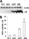
Increased SDS-soluble Aβ levels in the GRK5KO/APPsw mice. A, representative Western blots for SDS-soluble Aβ in the posterior cortex of WT, GRK5Het (KO), APPsw (APP), and the double heterozygote (DB) mice using mAb to Aβ clone 6E10. B, quantification of immunoblots for Aβ. The average densitometric values after β-actin normalization represent protein levels for each data point (n = 4). *, p < 0.05 versus APPsw mice.
FIGURE 4.
FIGURE 4.
Increased sAPPβ in the GRK5KO/APPsw mice. A, representative Western blots for full-length APP (fAPP) and sAPPβ in the posterior cortex of APPsw and double heterozygote mice (DB). B, quantification of immunoblots for full-length APP and sAPPβ. The average densitometric values after β-actin normalization represent protein levels for each data point (n = 6). *, p < 0.05 versus APPsw mice. AU, arbitrary units.
FIGURE 5.
FIGURE 5.
Dynamic changes of ISF Aβ in the young adult GRK5KO/APPsw mice. ISF Aβ in the microdialysates of hippocampus from the APPsw and the double heterozygote mice (DB) were collected in free-moving animals and measured using the human Aβ1–x ELISA kit, as detailed under “Experimental Procedures.” (n = 7). The basal ISF Aβ levels did not differ between the APPsw and the double heterozygote mice. After NOI, however, the ISF Aβ levels in the APPsw mice decreased over time and became significantly lower at 3 h after NOI. By contrast, the ISF Aβ levels in the double heterozygote mice barely decreased at all, indicating that the double heterozygote mice lost their ability to down-regulate the ISF Aβ in response to NOI stimulation. *, p < 0.05, as compared with either the basal level in the APPsw mice or the stimulated Aβ level at 3 h after NOI in the double mice.
FIGURE 6.
FIGURE 6.
Effects of MT and eserine on ISF Aβ in the young adult GRK5KO/APPsw mice. The treatment drugs were administered into the hippocampus via reverse microdialysis under NOI conditions, and the effects of eserine (A), MT (B), and their combination (C) on ISF Aβ in the young adult GRK5KO/APPsw mice were assessed as detailed under “Experimental Procedures.” A, treatment with eserine (1 μm) decreased the ISF Aβ significantly (*, p < 0.01 as compared with their own non-treated vehicle; n = 6) in both the APPsw and the double heterozygote mice but failed to correct the difference between the APPsw and the double (DB) mice. B, treatment with MT (1 μm) brought the ISF Aβ in the double heterozygote mice down to the level equivalent to that in the APPsw mice but did not significantly affect the ISF Aβ in the APPsw mice. Therefore, the MT treatment efficiently corrected the difference between the APPsw and the double heterozygote mice. n = 6, **, p < 0.001 as compared with vehicle; #, p < 0.001, for interactions between the MT treatment and the GRK5 deficiency analyzed using two-way analysis of variance. C, the combined effects of eserine and MT were larger than each treatment alone, but not significantly larger than their sum, indicating that these two treatments can additively work together to maximally decrease the ISF Aβ levels. n = 4, **, p < 0.001 as compared with vehicle.
FIGURE 7.
FIGURE 7.
Hypothetic model that links GRK5 deficiency to the amyloid and cholinergic hypotheses. This hypothetic model schematically illustrates the links of GRK5 deficiency to the amyloid and cholinergic hypotheses, with key references cited and abbreviations spelled out in this legend. The key pathologic molecule of the amyloid hypothesis, Aβ (1), is the main cause of GRK5 deficiency (11); the latter selectively impairs M2 receptor desensitization and leads to presynaptic M2 hyperactivity (17), which initiates cascading pathological events that ultimately result in selective cholinergic dysfunction and degeneration (12, 20–22), the key event of the cholinergic hypothesis (31). In brief, the presynaptic M2 hyperactivity has both anterograde and retrograde impact; anterogradely, it persistently inhibits ACh release, which causes postsynaptic cholinergic hypofunction, and retrogradely, it persistently suppresses cAMP-dependent signaling pathways, which, although inhibiting ACh release (41), may also promote cholinergic axonopathy and apoptosis. The cAMP-dependent signaling pathways have important roles in neuronal survival via 1) cAMP/PKA/CREB/Bcl-2 anti-apoptotic signaling (42–46), and 2) PKA also phosphorylates GSK3, including GSK3β, and inhibits their activities, cooperating with increased glucose hydrolysis for the increased energy consumption during neurite outgrowth (47–50). When these important signaling pathways are persistently suppressed by the presynaptic M2 hyperactivity, the cholinergic neurons may have disinhibited GSK3β, which increases Tau and KLC phosphorylation and contributes to axonopathy (51–55). Moreover, the inhibited anti-apoptotic pathway may also lead to increased vulnerability for the cholinergic neurons. On the anterograde direction, the postsynaptic cholinergic hypofunction, mainly mediated through M1 receptor (postsynaptic M1 hypofunction), leads to deficiencies in a number of important functions in cholinoceptive neurons, including: 1) memory deficiency (33); 2) decreased cAMP/PKA/CREB/Bcl-2 anti-apoptotic signaling strength (42–46, 56); 3) decreased cAMP/PKA signaling, which leads to decreased pGSK3β and increased GSK3β activity, as well as subsequently increased Tau and KLC phosphorylation (51–55, 57, 58); and 4) decreased activities of PKC and α-secretase (ADAM17/TACE) and increased β-secretase (BACE1) activity, which shifts APP processing in favor of β-amyloidogenesis (6, 57, 59). This last consequence of the cholinergic dysfunction leads to more Aβ production (as we demonstrated in this study), which pushes the GRK5 deficiency further and closes the loop of the vicious cycle between Aβ, GRK5 deficiency, and cholinergic dysfunction. Of course, more Aβ production during aging means more Aβ deposits, inflammation, and non-selective toxic effects. All these toxic degenerative insults, along with increased free radicals during aging, will come back to attack the already vulnerable cholinergic system, eventually leading to the cholinergic selective neurodegeneration in AD. Abbreviations used are: acetyl-CoA, acetyl-coenzyme A; ADAM17, a disintegrin and metalloproteinase domain 17; BACE1, β-site of APP-cleaving enzyme 1; fAβ, fibrillar Aβ; pCREB, phosphorylated cAMP response element-binding protein; pGSK3β, phosphorylated glycogen synthase kinase 3β; PKA, cAMP-dependent protein kinase; PKC, protein kinase C; P-KLC, phosphorylated KLC; P-Tau, phosphorylated Tau; TACE, tumor necrosis factor-α-converting enzyme; VAChT, vesicular ACh transporter.

Similar articles

Cited by

References

    1. Hardy J., Selkoe D. J. (2002) Science 297, 353–356 - PubMed
    1. Tanzi R. E., Bertram L. (2005) Cell 120, 545–555 - PubMed
    1. Roberson M. R., Harrell L. E. (1997) Brain Res. Brain Res. Rev. 25, 50–69 - PubMed
    1. Lee R. K., Wurtman R. J., Cox A. J., Nitsch R. M. (1995) Proc. Natl. Acad. Sci. U.S.A. 92, 8083–8087 - PMC - PubMed
    1. Nitsch R. M., Deng M., Growdon J. H., Wurtman R. J. (1996) J. Biol. Chem. 271, 4188–4194 - PubMed

Publication types