. 2010 Nov 1;24(21):2383-8.
doi: 10.1101/gad.1978810.

The Hippo signaling pathway restricts the oncogenic potential of an intestinal regeneration program

Affiliations

The Hippo signaling pathway restricts the oncogenic potential of an intestinal regeneration program

Jing Cai et al. Genes Dev. .

Abstract

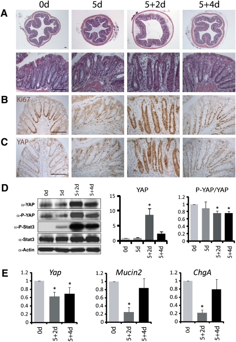
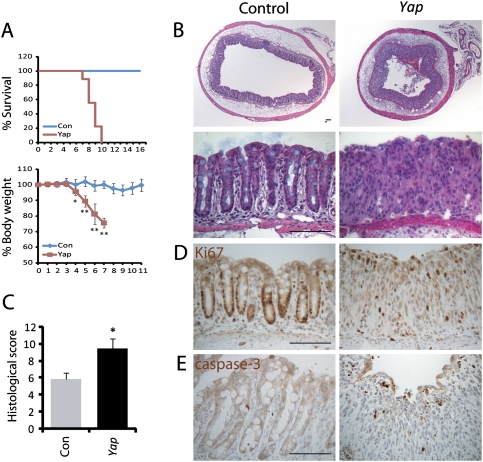
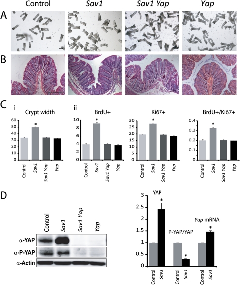
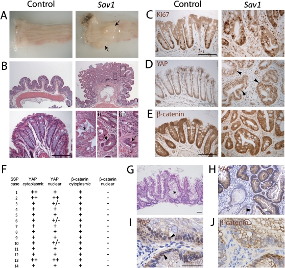
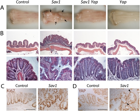
Although a developmental role for Hippo signaling in organ size control is well appreciated, how this pathway functions in tissue regeneration is largely unknown. Here we address this issue using a dextran sodium sulfate (DSS)-induced colonic regeneration model. We find that regenerating crypts express elevated Yes-associated protein (YAP) levels. Inactivation of YAP causes no obvious intestinal defects under normal homeostasis, but severely impairs DSS-induced intestinal regeneration. Conversely, hyperactivation of YAP results in widespread early-onset polyp formation following DSS treatment. Thus, the YAP oncoprotein must be exquisitely controlled in tissue regeneration to allow compensatory proliferation and prevent the intrinsic oncogenic potential of a tissue regeneration program.


Figures

Figure 1.
Figure 1.
Increased YAP protein levels in regenerating crypts. (A) H&E staining of colon sections from 8-wk-old wild-type mice before (0 d) and after a 5-d DSS treatment (5 d), followed by normal drinking water for 2 d (5 + 2 d) or 4 d (5 + 4 d). (Top row) Low magnification. (Bottom row) High magnification. (B) Ki67 staining. Note the restriction of Ki67-positive proliferating cells to the crypt base in a normal colon, the loss of Ki67-positive cells in 5-d crypts, the expansion of Ki67-positive cells from the crypt base to the whole regenerating crypt in 5 + 2-d colons, and the restoration of Ki67-positive cells to the crypt base in 5 + 4-d colons. (C) YAP staining. In wild-type crypts, YAP was detected in the entire crypt epithelium. Note the dramatic increase of YAP staining in 5 + 2-d crypts. Also note the absence of appreciable YAP nuclear accumulation in control and regenerating crypts. (D) Western blotting analysis. Protein extracts from control and regenerating crypts were probed with the indicated antibodies. Note the increase of YAP, P-YAP, and P-Stat3 in 5 + 2-d and 5 + 4-d crypts. The YAP protein level and P-YAP/YAP ratio were quantified in the graphs to the right. Data are mean ± SD. n = 3. (*) P < 0.01, t-test. (E) Real-time PCR analysis. mRNAs from control and regenerating crypts were analyzed for the expression of the indicated genes. Note the decrease of Yap mRNA in 5 + 2-d and 5 + 4-d crypts. Also note the decreased expression of the goblet cell marker Mucin2 and the enteroendocrine cell marker Chromogranin A (ChgA) in 5 + 2-d crypts, and the recovery of their expression levels in 5 + 4-d crypts. Data are mean ± SD. n = 3. (*) P < 0.01, t-test. Bars, 100 μm.
Figure 2.
Figure 2.
Impaired regeneration of Yap-deficient colonic crypts. (A) Increased mortality and loss of body weight in Yap-deficient mice after DSS treatment. Mice were treated with 2.5% DSS for 7 d and supplied with normal drinking water thereafter. Thirteen wild-type and nine Yap-deficient mice were treated for mortality rate analysis. Nine wild-type and eight Yap-deficient mice were treated for body weight analysis. (*) P < 0.05; (**) P < 0.01, t-test. (B) H&E staining of colon sections after a 7-d DSS treatment. (Top row) Low magnification. (Bottom row) High magnification. Note the absence of crypt structure and the expansion of stromal cells in Yap-deficient colons. (C) Histological score of colons after a 7-d DSS treatment. Tissue damages were quantified as described in the Materials and Methods. Eight wild-type and nine Yap-deficient mice were treated for this analysis. Data are mean ± SEM. (*) P < 0.05, t-test. (D,E) Ki67 and cleaved caspase-3 staining showing fewer proliferating and more apoptotic cells in Yap-deficient crypts after DSS treatment. Bars, 100 μm.
Figure 3.
Figure 3.
Loss of Sav1 results in Yap-dependent crypt hyperplasia. (A) Isolated colonic crypts from 4-wk-old wild-type, Sav1, Yap, or Sav1 Yap double-mutant colons. Note the enlarged width of Sav1 mutant crypts. Quantification of crypt width is shown in C, graph i. (B) H&E staining of the colonic sections from 4-wk-old wild-type, Sav1, Yap, or Sav1 Yap double-mutant colons. Note the enlarged width of Sav1 mutant crypts. (C, graph i) Quantification of crypt width from A. Eighty crypts from three mice of each genotype were used. Data are mean ± SEM. (*) P < 0.01, t-test. (Graph ii) Quantification of cell proliferation. Four-week-old wild-type, Sav1, Yap, or Sav1 Yap double-mutant mice were analyzed for BrdU and Ki67 staining 2 h after a single i.p. injection of BrdU. The number of BrdU+ or Ki67+ cells and the ratio of BrdU+/Ki67+ in each crypt were quantified with 200 crypts from three mice of each genotype. Data are mean ± SEM. (*) P < 0.01, t-test. (D) Quantification of YAP protein and mRNA levels. Note the increased YAP protein and mRNA levels in Sav1-deficient crypts. Also note that, while the absolute amount of P-YAP was similar in wild-type and Sav1-deficient crypts, the P-YAP/YAP ratio was decreased in Sav1-deficient crypts. Data used in the graph are mean ± SD. n = 3. (*) P < 0.01, t-test. Bars, 100 μm.
Figure 4.
Figure 4.
SSP development in Sav1-deficient mouse colons and the prevalence of Hippo pathway dysregulation in human SSPs. (A) Distal colon of 13-mo-old wild-type control and Sav1-deficient littermates. Note the presence of two polyps in the Sav1-deficient colon (black arrows). (B) H&E staining of the colonic sections from 13-mo-old wild-type control (left) and Sav1-deficient (right) littermates. The top and bottom panels show the corresponding low- and high-magnification images, respectively. Note the presence of serrated polyps with “saw-tooth” crypt epithelium (asterisk in panel i), adenomatous transformation (arrow in panel ii), and invasion of the muscularis mucosa (arrow in panel iii) in Sav1-deficient colons. (C) Ki67 staining. Note that Ki67-positive proliferating cells were restricted to the crypt base in control colons. In Sav1-deficient colons, scattered Ki67-positive cells were detected throughout the crypt epithelia. (D,E) YAP and β-catenin staining. Note the accumulation of nuclear YAP but not nuclear β-catenin in Sav1-deficient colonic polyps (arrows). (F–J) Analysis of human SSPs. (F) Summary of YAP and β-catenin staining in human SSPs. (G) Histopathology of a right-sided SSP obtained by endoscopic excision. H&E stain, 10× magnification. Note the characteristic polypoid epithelium. (H) Up-regulation of YAP in the polypoid epithelium of an SSP compared with adjacent normal colonic epithelium. At the bottom right half of the panel, one gland was partially involved by SSP and demonstrated YAP up-regulation (white arrowhead), while the nonneoplastic half of the gland had minimal expression (black arrowhead). (I) High-power magnification of SSP epithelium with nuclear YAP labeling. Magnification, 40×. (J) Absence of nuclear β-catenin, with retained membranous localization of the protein, in the serial section of the SSP shown in D. Magnification, 40×. Bars, 100 μm.
Figure 5.
Figure 5.
DSS-induced regeneration accelerated polyp development in Sav1-deficient colons in a Yap-dependent manner. (A) Distal colon of 12-wk-old wild-type, Sav1, Yap, or Sav1 Yap double-mutant mice treated with 2.5% DSS for 4 d, followed by normal drinking water for 3 mo. Note the presence of multiple large colonic polyps in the Sav1-deficient colon (arrows). (B) H&E staining of colonic sections from animals in A. The top and bottom panels show the corresponding low- and high-magnification images, respectively. Note the presence of serrated crypt epithelium in Sav1-deficient polyps (asterisk). (C) Ki67 staining of colon sections from control and Sav1-deficient littermates. Note the presence of scattered Ki67-positive cells throughout the serrated crypt epithelium. (D) YAP staining of colon sections from control and Sav1-deficient littermates. Note the accumulation of nuclear YAP in Sav1-deficient colonic polyps (arrowheads). Bars, 100 μm.

Comment in

  • YAP tips the balance.
    Skinner M. Skinner M. Nat Rev Cancer. 2010 Dec;10(12):811. doi: 10.1038/nrc2973. Nat Rev Cancer. 2010. PMID: 21155175 No abstract available.

References

    1. Badouel C, Garg A, McNeill H 2009. Herding Hippos: Regulating growth in flies and man. Curr Opin Cell Biol 21: 837–843 - PubMed
    1. Beachy PA, Karhadkar SS, Berman DM 2004. Tissue repair and stem cell renewal in carcinogenesis. Nature 432: 324–331 - PubMed
    1. Bollrath J, Phesse TJ, von Burstin VA, Putoczki T, Bennecke M, Bateman T, Nebelsiek T, Lundgren-May T, Canli O, Schwitalla S, et al. 2009. gp130-mediated Stat3 activation in enterocytes regulates cell survival and cell-cycle progression during colitis-associated tumorigenesis. Cancer Cell 15: 91–102 - PubMed
    1. Camargo FD, Gokhale S, Johnnidis JB, Fu D, Bell GW, Jaenisch R, Brummelkamp TR 2007. YAP1 increases organ size and expands undifferentiated progenitor cells. Curr Biol 17: 2054–2060 - PubMed
    1. Dong J, Feldmann G, Huang J, Wu S, Zhang N, Comerford SA, Gayyed MF, Anders RA, Maitra A, Pan D 2007. Elucidation of a universal size-control mechanism in Drosophila and mammals. Cell 130: 1120–1133 - PMC - PubMed

Publication types

MeSH terms

LinkOut - more resources