. 2010 Nov 1;24(21):2420-9.
doi: 10.1101/gad.1954410.

The Lats2 tumor suppressor augments p53-mediated apoptosis by promoting the nuclear proapoptotic function of ASPP1

Affiliations

The Lats2 tumor suppressor augments p53-mediated apoptosis by promoting the nuclear proapoptotic function of ASPP1

Yael Aylon et al. Genes Dev. .

Abstract

Apoptosis is an important mechanism to eliminate potentially tumorigenic cells. The tumor suppressor p53 plays a pivotal role in this process. Many tumors harbor mutant p53, but others evade its tumor-suppressive effects by altering the expression of proteins that regulate the p53 pathway. ASPP1 (apoptosis-stimulating protein of p53-1) is a key mediator of the nuclear p53 apoptotic response. Under basal conditions, ASPP1 is cytoplasmic. We report that, in response to oncogenic stress, the tumor suppressor Lats2 (large tumor suppressor 2) phosphorylates ASPP1 and drives its translocation into the nucleus. Together, Lats2 and ASPP1 shunt p53 to proapoptotic promoters and promote the death of polyploid cells. These effects are overridden by the Yap1 (Yes-associated protein 1) oncoprotein, which disrupts Lats2-ASPP1 binding and antagonizes the tumor-suppressing function of the Lats2/ASPP1/p53 axis.


Figures
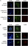
Figure 1.
Figure 1.
ASPP1 is translocated from the cytoplasm to the nucleus in a Lats2-dependent manner. (A) WI-38 cells were retrovirally infected with H-RasV12 or empty vector only. Two days after infection, cells were transfected with Lats2 siRNA (siLats2) or control siRNA (siControl). Three days after infection, cells were fixed and immunostained with the LA2 antibody (Toji et al. 2004) to visualize endogenous Lats2, and with anti-ASPP1 antibodies. Nuclear DNA was visualized by DAPI staining. (B) HCT116 cells were transiently transfected with V5-tagged ASPP1 together with either empty vector, kinase-active myc-tagged Lats2 (Lats2), or kinase-dead myc-tagged Lats2 (Lats2-KD). ASPP1 and Lats2 were detected with anti-V5 and anti-Lats2 antibodies, respectively. Nuclear DNA was visualized by DAPI staining.
Figure 2.
Figure 2.
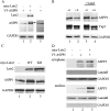
Lats2 phosphorylates ASPP1. (A) HCT116 cells were transiently transfected with V5-tagged ASPP1 together with empty vector or myc-tagged Lats2. Cells were harvested 24 h after transfection, and lysates were subjected to Western blot analysis. Lats2 and ASPP1 were detected by anti-myc tag and anti-V5 antibodies, respectively. GAPDH was used as a loading control. Lane 3 represents a longer exposure of lane 1. (B) HCT116 cells were transiently transfected as in A. Cell lysates were either mock-treated or treated with λ phosphatase (λ). Equivalent amounts were analyzed by Western blot. Ablation of phospho-Yap1 was used as a positive control for phosphatase activity. GAPDH served as a loading control. (C) HCT116 cells were transiently transfected as in A with empty vector, myc-tagged wild-type Lats2 (WT), or myc-tagged kinase-dead Lats2 (KD). GAPDH served as a loading control. (D) HCT116 cells were transiently transfected as in A. Twenty-four hours after transfection, cell lysates were processed for nuclear and cytoplasmic fractions. Equivalent amounts of each fraction were analyzed by Western blot. LaminB and GAPDH were used to verify the purity of the nuclear and cytoplasmic fractions, respectively.
Figure 3.
Figure 3.
Lats2 and ASPP1 interact within cells. (A) Schematic representation of full-length ASPP1 and different deletion mutants lacking either the N terminus (ΔN), the ANK repeat (ANK), and SH3 domain (ΔC), or the SH3 domain only (small ΔC). (B) HCT116 cells were transiently transfected with V5-tagged full-length ASPP1 or deletion mutants thereof, together with empty vector or myc-tagged Lats2. Lysates were immunoprecipitated (IP) with either anti-V5 or anti-myc antibodies. Westerns were immunoblotted (IB) using antibodies directed against either V5 tag, myc tag, or GAPDH. (DW) Direct Western: 2.5% of each lysate subjected directly to SDS-PAGE and Western blot analysis. (C) WI-38 cells were infected with H-RasV12 or empty retroviral vector. Two days after infection, cells were harvested and subjected to immunoprecipitation with either anti-Lats2, anti-ASPP1, or control anti-HA antibodies. Membranes were immunoblotted (IB) using anti-ASPP1, anti-Lats2 (LA2), or anti-GAPDH antibodies. Direct Western (DW) is as in B.
Figure 4.
Figure 4.
Lats2 and ASPP1 shift p53 binding toward proapoptotic promoters. (A) HCT116 cells were transiently transfected with vector or H-RasV12. Twenty-four hours after transfection, cell lysates were separated into chromatin-bound and chromatin-unbound fractions. Equivalent amounts of each fraction were analyzed by Western blot using antibodies against ASPP1, Lats2, and p53. H2B and GAPDH were used to verify the purity of the bound and unbound fractions, respectively. (B) HCT116 cells were transiently transfected with the indicated combinations of V5-tagged ASPP1 and myc-tagged Lats2. Forty-eight hours later, ChIP was performed using antibodies against V5 tag to immunoprecipitate ASPP1. Input and bound DNA were quantified using qPCR with primer pairs corresponding to p53-responsive elements within the indicated genes. (C) HCT116 cells were transiently transfected with Flag-tagged H2B together with the indicated combinations of vector, myc-tagged Lats2, and H-RasV12. Lysates were immunoprecipitated (IP) using anti-Flag antibodies and then eluted using Flag peptide. Westerns were immunoblotted (IB) using anti-myc or anti-Flag antibodies to detect Lats2 or H2B, respectively. Direct Western (DW) is as in Figure 3B. (D,E) ChIP was performed as in B, except that anti-myc tag or anti-p53 (CM1) antibodies were used to precipitate Lats2 or p53, respectively.
Figure 5.
Figure 5.
Lats2 and ASPP1 modulate p53 target gene preference. (A) HCT116 cells were transiently transfected with combinations of vector, ASPP1, and Lats2 together with the indicated firefly luciferase reporter plasmids. Twenty-four hours later, cells were either mock-treated or treated with 5-FU (50 μg/mL) for 16 h. Luciferase assays were performed in triplicate, and values represent average firefly luciferase expression after normalization for cotransfected renilla luciferase plasmid. (B) WI-38 cells were infected with H-RasV12 or empty retroviral vector. Two days later, cells were transiently transfected with siRNA oligonucleotides specific for ASPP1 or Lats2 or control siRNA (siASPP1, siLats2, and siCont, respectively). Three days after infection, cells were harvested, and RNA was extracted and subjected to qRT–PCR analysis. Values were normalized to HPRT mRNA.
Figure 6.
Figure 6.
Lats2 and ASPP1 promote apoptosis of polyploid cells. (A) WI-38 cells were infected with H-RasV12 or empty retroviral vector. Two days after infection, cells were transfected with siRNA oligonucleotides specific for ASPP1 or Lats2 or control siRNA (siASPP1, siLats2, and siCont, respectively). Three days after infection, cells were harvested, fixed, stained with PI, and subjected to FACS-based DNA content analysis. (B) Graphic representation of averaged values corresponding to the indicated cell subpopulations in A. (C) HCT116 cells were transiently transfected with GFP-tagged Lats2, together with the indicated combinations of H-RasV12 and Flag-tagged Yap1. Twenty-four hours later, cells were fixed and subjected to analysis by light microscopy. Nuclear DNA was detected by DAPI staining. Single representative cells are magnified and presented in the far right panel. (D) HCT116 cells were transiently transfected with V5-tagged ASPP1, myc-tagged Lats2, and Flag-tagged Yap1. Twenty-four hours later, cells were fixed and immunostained using anti-V5 or anti-Flag antibodies. Nuclear DNA was detected by DAPI staining. Cells marked 1 and 2 are representative examples of nuclear and cytoplasmic ASPP1 staining, respectively.
Figure 7.
Figure 7.
Yap1 antagonizes the effects of Lats2 and ASPP1. (A) HCT116 cells were transiently transfected with combinations of V5-tagged ASPP1 (0.5 μg [+] or 2 μg [++]), Flag-tagged Yap1 (0.5 μg [+] or 2 μg [++]), and myc-tagged Lats2 (3 μg [+]). Anti-myc tag antibodies were used to coimmunoprecipitate Lats2. Western blot analysis was performed; Lats2, ASPP1, and Yap1 were visualized using anti-myc tag, anti-V5 tag, and anti-Flag antibodies, respectively. Direct Western (DW) is as in Figure 3B. GAPDH was used as a loading control. (B) HCT116 cells were transiently transfected with combinations of siRNA oligonucleotides specific for Yap1 and ASPP1, or control siRNA (siYap1, siASPP1, and siCont, respectively). Twenty-four hours later, cells were either mock-treated or treated with 5-FU (50 μg/mL) for 16 h. Cells were then harvested, and RNA was extracted and subjected to qRT–PCR analysis. Values were normalized to HPRT mRNA. (C) Proposed model for cross-talk between oncogenic Ras, Lats2, APP1, Yap1, and p53.

Comment in

  • YAP tips the balance.
    Skinner M. Skinner M. Nat Rev Cancer. 2010 Dec;10(12):811. doi: 10.1038/nrc2973. Nat Rev Cancer. 2010. PMID: 21155175 No abstract available.

Similar articles

Cited by

References

    1. Agirre X, Roman-Gomez J, Jimenez-Velasco A, Garate L, Montiel-Duarte C, Navarro G, Vazquez I, Zalacain M, Calasanz MJ, Heiniger A, et al. 2006. ASPP1, a common activator of TP53, is inactivated by aberrant methylation of its promoter in acute lymphoblastic leukemia. Oncogene 25: 1862–1870 - PubMed
    1. Aylon Y, Michael D, Shmueli A, Yabuta N, Nojima H, Oren M 2006. A positive feedback loop between the p53 and Lats2 tumor suppressors prevents tetraploidization. Genes Dev 20: 2687–2700 - PMC - PubMed
    1. Aylon Y, Yabuta N, Besserglick H, Buganim Y, Rotter V, Nojima H, Oren M 2009. Silencing of the Lats2 tumor suppressor overrides a p53-dependent oncogenic stress checkpoint and enables mutant H-Ras-driven cell transformation. Oncogene 28: 4469–4479 - PMC - PubMed
    1. Bergamaschi D, Samuels Y, O'Neil NJ, Trigiante G, Crook T, Hsieh JK, O'Connor DJ, Zhong S, Campargue I, Tomlinson ML, et al. 2003. iASPP oncoprotein is a key inhibitor of p53 conserved from worm to human. Nat Genet 33: 162–167 - PubMed
    1. Chen CF, Yeh SH, Chen DS, Chen PJ, Jou YS 2005. Molecular genetic evidence supporting a novel human hepatocellular carcinoma tumor suppressor locus at 13q12.11. Genes Chromosomes Cancer 44: 320–328 - PubMed

Publication types

MeSH terms