. 2010 Nov 1;24(21):2430-9.
doi: 10.1101/gad.1954310.

Cytoplasmic ASPP1 inhibits apoptosis through the control of YAP

Affiliations

Cytoplasmic ASPP1 inhibits apoptosis through the control of YAP

Arnaud M Vigneron et al. Genes Dev. .

Abstract

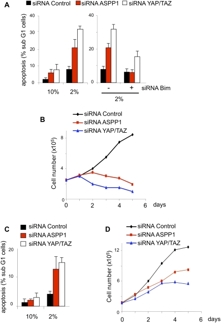
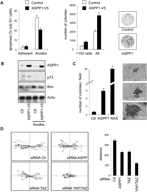
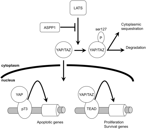
The ASPP (apoptosis-stimulating protein of p53) family of proteins can function in the nucleus to modulate the transcriptional activity of p53, with ASPP1 and ASPP2 contributing to the expression of apoptotic target genes. In this study, we describe a new function for cytoplasmic ASPP1 in controlling YAP (Yes-associated protein)/TAZ. ASPP1 can inhibit the interaction of YAP with LATS1 (large tumor suppressor 1), a kinase that phosphorylates YAP/TAZ and promotes cytoplasmic sequestration and protein degradation. This function of ASPP1 therefore enhances nuclear accumulation of YAP/TAZ and YAP/TAZ-dependent transcriptional regulation. The consequence of YAP/TAZ activation by ASPP1 is to inhibit apoptosis, in part through the down-regulation of Bim expression, leading to resistance to anoikis and enhanced cell migration. These results reveal a potential oncogenic role for cytoplasmic ASPP1, in contrast to the tumor-suppressive activity described previously for nuclear ASPP1.


Figures

Figure 1.
Figure 1.
Cytoplasmic ASPP1 regulates gene expression. (A) Nuclear and cytoplasmic expression of ASPP1 in different cell lines detected by Western blot in control conditions or after treatment for 24 h with 2 mM HU. Lamin expression was used as loading and purity control for fractionation. (B) HCT116 cells were transfected with a pool of siRNA against ASPP1 or control siRNA, and ASPP1 expression was assessed by Western blot in control conditions or after 2 mM HU or 10 μM Nutlin treatment. (C) mRNA expression of the indicated genes was then determined by RT-qPCR with specific primers. The results were normalized against two different standard genes, and the graphs represent the mean of five independent experiments. (D) Expression of Pig3, Puma, and Bax assessed by Western blot in U2OS cells treated with the indicated siRNAs with or without 10 μM Nutlin for 30 h. Actin expression was used as a loading control. (E) mRNA expression of 130 genes quantified by RT-qPR in the same conditions as in B, shown as a dot plot. Results following control siRNA treatment are shown in the X-axis, and ASPP1 siRNA treatment is shown in the Y-axis. The YAP/TAZ target genes are highlighted by a red X.
Figure 2.
Figure 2.
ASPP1 controls YAP target genes. (A) CTGF, PHLDB2, and Bim mRNA expression measured by RT-qPCR with specific primer in HCT116 cells transfected with control or ASPP1 siRNA, and treated or not treated with 400 μM HU. The results were normalized against two different standard genes, and the graphs represent the mean of five independent experiments. (B) Expression of YAP, TAZ, and Bim assessed by Western blot in HCT116 cells treated with the indicated siRNAs, with or without 400 μM HU, for 24 h. (C) Same experiment as in A in the HCT116 p53−/− cell line. (D) Apoptosis in HCT116p53−/− cells was quantified under the same conditions as in C by DNA staining and flow cytometry analysis. The graph is representative of three independent experiments and shows the percentage of cell in the sub-G1 fraction.
Figure 3.
Figure 3.
ASPP1 increases YAP and TAZ nuclear localization. (A) Immunofluorescence staining of endogenous YAP/TAZ in U2OS cells infected with a control or ASPP1 expression vector. Cells were counterstained with DAPI to localize the presence of YAP/TAZ in the nucleus. More than 10,000 cells were analyzed using an Operetta screening system, and the graph represents the percentage of cells expressing YAP/TAZ in the cytoplasm in three different experiments. ASPP1 overexpression was confirmed by immunoblot using actin as a loading control. (B) The same experiment as in A repeated in HCT116 transfected with control or ASPP1 siRNA. The graph represents the percentage of cells expressing YAP/TAZ in the cytoplasm in three different experiments. (C) YAP Ser127 phosphorylation analyzed by Western blot in U2OS cells transiently transfected with a YAP expression vector and an increasing concentration of a vector coding for ASPP1. (D) YAP Ser127 phosphorylation analysis by Western blot in HCT116 cells transfected with control or ASPP1 siRNA, with or without HU. The loading was normalized for total YAP to allow a comparison of the phosphorylation levels. (E) Unbound and chromatin-bound YAP and TAZ was fractionated by detergent and DNase treatment in HCT116 cells under the same conditions as in D. YAP phosphorylated on Ser127, total YAP, and TAZ levels were assessed by immunoblot. The high-mobility group protein HMGA2 was used as a control for the chromatin-bound fraction.
Figure 4.
Figure 4.
Stabilization of YAP and TAZ proteins in response to ASPP1. (A) Expression of YAP and ASPP1 assessed by Western blot or RT-qPCR in HCT116 cells treated with control or ASPP1 siRNA, with or without 400 μM HU. Actin was used as a loading control. mRNA expression quantification was normalized against two different standard genes and represents five independent experiments. (B) YAP protein stability was tested in U2OS cells infected with a control or ASPP1 coding vector and treated with cyclohexamide for the indicated time. YAP expression was measured by Western blot using actin as a loading control. The graph represents YAP protein expression normalized to its initial level, and represents the mean of three independent experiments. (C) TAZ protein stability was tested in U2OS under the same conditions as described in B.
Figure 5.
Figure 5.
ASPP1–LATS1 interaction impedes LATS1–YAP complex formation. (A) Endogenous ASPP1 was immunoprecipitated from HCT116 cells. The immunoprecipitation was then resolved by SDS-PAGE and immunoblotted for LATS1, LATS2, YAP, and ASPP1. Five percent of the lysate was loaded onto the gel to assess input protein levels. (B) U2OS cells were transiently transfected with vectors encoding LATS1 and YAP and an increasing concentration of a vector encoding ASPP1. LATS1 was then immunoprecipitated, resolved by SDS-PAGE, and immunoblotted for LATS1, ASPP1, and YAP. Five percent of the lysate was loaded onto the gel to assess the input levels of ASPP1 and YAP. (C) Same experiment as in B examining LATS2. (D) Diagram showing the different domains of LATS1. (E) U2OS cells infected with ASPP1 were transiently transfected with vectors encoding Myc-tagged wild-type or mutant LATS1 as indicated. ASPP1 immunoprecipitations were resolved by SDS-PAGE and immunoblotted for ASPP1 using the anti-Myc antibody. Ten percent of the lysate was loaded onto the gel to assess the input levels of the different LATS1 constructs. (F) Immunofluorescence staining of ASPP1 and LATS1 expressed in U2OS cells, showing the colocalization of ASPP1 with wild-type LATS1 but not LATS-ΔKD. (G) Endogenous YAP was immunoprecipitated from HCT116 cells treated with control or YAP siRNAs. The immunoprecipitation was then resolved by SDS-PAGE and immunoblotted for LATS1 and LATS2. Five percent of the lysate was loaded onto the gel to assess input protein levels. (H) Endogenous YAP was immunoprecipitated from HCT116 cells treated with control or ASPP1 siRNAs. The immunoprecipitation was then resolved by SDS-PAGE and immunoblotted for LATS1 and YAP. Five percent of the lysate was loaded onto the gel to assess input protein levels.
Figure 6.
Figure 6.
ASPP1 and YAP inhibit apoptosis under low-serum conditions. (A) HCT116 cells were transfected with different combinations of control siRNA or siRNAs directed against ASPP1, YAP, and Bim. Cells were then incubated for 48 h in 10% or 2% serum and apoptosis was measured by the analysis of the sub-G1 fraction. The graph represents the mean of three independent experiments. (B) HCT116 cells were plated at low confluence and transfected with control, ASPP1, or YAP siRNAs. Cells were maintained in 2% serum and harvested each day for 6 d. Live cells were counted by Trypan blue exclusion; the graph represents the mean of three independent experiments. (C) Same experiments as in A in the HCT116 p53−/− cell line. (D) Same experiments as in B in the HCT116 p53−/− cell line.
Figure 7.
Figure 7.
ASPP1 protects cells from anoikis. (A) U2OS cells infected with a control or ASPP1 coding vector were incubated for 4 d in a dish coated with 1% agarose. Apoptosis induction was then measured by DNA staining and flow cytometry. The graph is representative of three independent experiments and shows the percentage of cells in the sub-G1 fraction. Alternatively, surviving cells were replated in normal conditions to allow them to recover from the matrix detachment period. Colonies formed 1 wk later were stained with Giemsa and quantified with ImageJ software. The graph shows the mean of three different experiments, and a dish from each condition is shown as an example. (B) p73, Bim, and ASPP1 expression in U2OS cells treated in the same way as in A were assessed by Western blot. Actin was used as a loading control. (C) Anchorage-independent growth of U2OS cells infected with a control or ASPP1 retroviral vector. Cells were embedded in 0.3% agar and left growing for 2 wk. The number of colonies was counted in a minimum of 50 different microscopic fields per condition and plotted as a mean of colonies per field. RAS-overexpressing U2OS cells were used as a positive control, and results show the mean of three different experiments. A representative colony for each condition is shown: for ASPP1- and RAS-expressing cells, an average-sized colony is shown; for the control cells, the largest colony seen is shown. (D) HCT116 cells transfected with control, ASPP1, TAZ, or YAP siRNA were observed by time-lapse video microscopy, and the movement of individual cells was followed using cell-tracking software. The results are presented as overlays of representative trajectories described by cells during their migration into the wound. The total distance of migration was extracted from the track plots. Values are mean ± SEM of >100 track plots from three independent experiments.
Figure 8.
Figure 8.
Model of YAP/TAZ regulation by cytoplasmic ASPP1. Under normal conditions, the activation of LATS leads to the phosphorylation of YAP/TAZ and their inactivation through cytoplasmic sequestration and degradation. ASPP1 can function to inhibit this activity of LATS1, allowing the nuclear translocation of YAP/TAZ where they can regulate gene expression. While this study shows an effect of ASPP1 on the regulation of YAP/TAZ-mediated survival signaling, it is also possible that ASPP1 can modulate the induction of apoptotic target gene expression regulated by a YAP/p73 complex.

Comment in

  • YAP tips the balance.
    Skinner M. Skinner M. Nat Rev Cancer. 2010 Dec;10(12):811. doi: 10.1038/nrc2973. Nat Rev Cancer. 2010. PMID: 21155175 No abstract available.

Similar articles

Cited by

References

    1. Agirre X, Roman-Gomez J, Jimenez-Velasco A, Garate L, Montiel-Duarte C, Navarro G, Vazquez I, Zalacain M, Calasanz MJ, Heiniger A, et al. 2006. ASPP1, a common activator of TP53, is inactivated by aberrant methylation of its promoter in acute lymphoblastic leukemia. Oncogene 25: 1862–1870 - PubMed
    1. Ahn J, Byeon IJ, Byeon CH, Gronenborn AM 2009. Insight into the structural basis of pro- and antiapoptotic p53 modulation by ASPP proteins. J Biol Chem 284: 13812–13822 - PMC - PubMed
    1. Aylon Y, Michael D, Shmueli A, Yabuta N, Nojima H, Oren M 2006. A positive feedback loop between the p53 and Lats2 tumor suppressors prevents tetraploidization. Genes Dev 20: 2687–2700 - PMC - PubMed
    1. Aylon Y, Ofir-Rosenfeld Y, Yabuta N, Lapi E, Nojima H, Lu X, Oren M 2010. The Lats2 tumor suppressor augments p53-mediated apoptosis by promoting the nuclear proapoptotic function of ASPP1. Genes Dev (this issue) doi: 10.1101/gad.1954410 - PMC - PubMed
    1. Bergamaschi D, Samuels-Lev Y, O'Neil NJ, Trigiante G, Crook T, Hseih JK, O'Conner DJ, Zhong S, Compargue I, Tomlinson ML, et al. 2003. iASPP oncoprotein is a key inhibitor of p53 conserved from worms to humans. Nat Genet 33: 162–167 - PubMed

Publication types

MeSH terms