. 2010 Nov 1;191(3):661-75.
doi: 10.1083/jcb.201007086.

The Cdc42-selective GAP rich regulates postsynaptic development and retrograde BMP transsynaptic signaling

Affiliations

The Cdc42-selective GAP rich regulates postsynaptic development and retrograde BMP transsynaptic signaling

Minyeop Nahm et al. J Cell Biol. .

Abstract

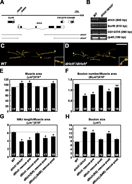
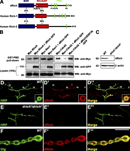
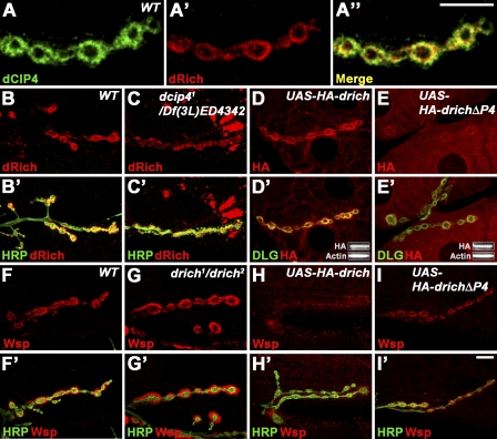
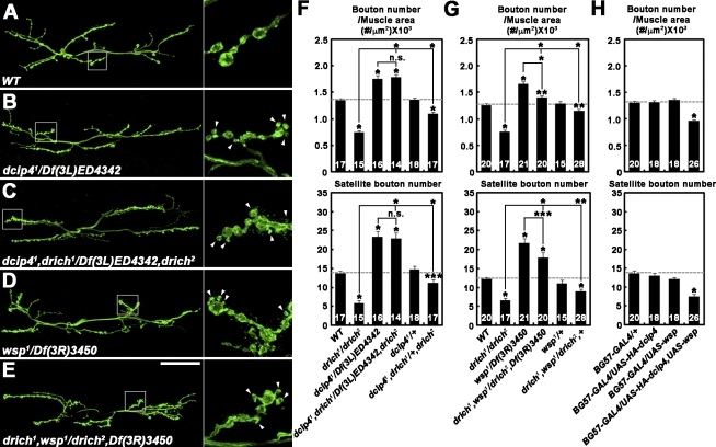
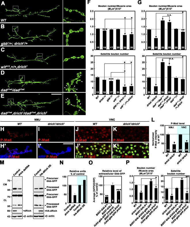
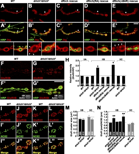
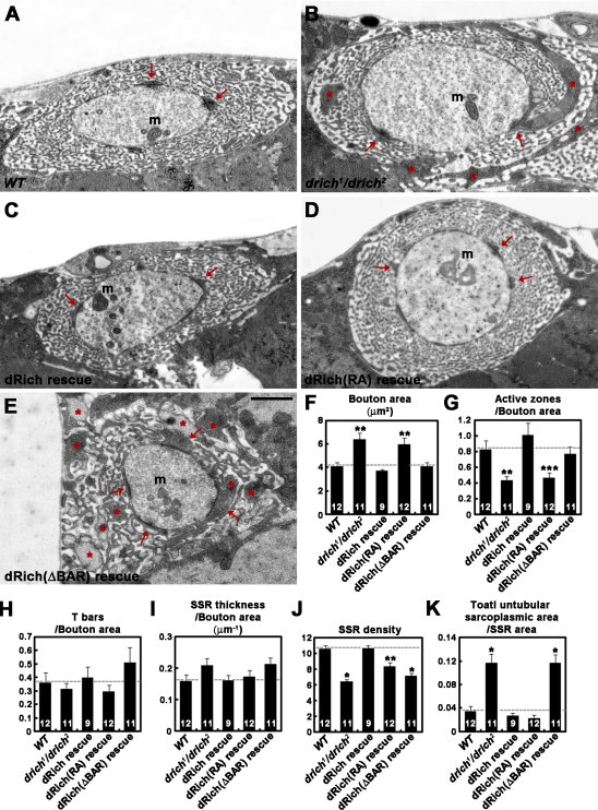
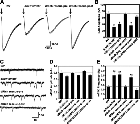
Retrograde bone morphogenetic protein signaling mediated by the Glass bottom boat (Gbb) ligand modulates structural and functional synaptogenesis at the Drosophila melanogaster neuromuscular junction. However, the molecular mechanisms regulating postsynaptic Gbb release are poorly understood. In this study, we show that Drosophila Rich (dRich), a conserved Cdc42-selective guanosine triphosphatase-activating protein (GAP), inhibits the Cdc42-Wsp pathway to stimulate postsynaptic Gbb release. Loss of dRich causes synaptic undergrowth and strongly impairs neurotransmitter release. These presynaptic defects are rescued by targeted postsynaptic expression of wild-type dRich but not a GAP-deficient mutant. dRich inhibits the postsynaptic localization of the Cdc42 effector Wsp (Drosophila orthologue of mammalian Wiskott-Aldrich syndrome protein, WASp), and manifestation of synaptogenesis defects in drich mutants requires Wsp signaling. In addition, dRich regulates postsynaptic organization independently of Cdc42. Importantly, dRich increases Gbb release and elevates presynaptic phosphorylated Mad levels. We propose that dRich coordinates the Gbb-dependent modulation of synaptic growth and function with postsynaptic development.


Figures

Figure 1.
Figure 1.
dRich required postsynaptically for presynaptic growth. (A) Genomic organization of drich/RhoGAP92B locus. The exon–intron organization of drich and neighboring genes Surf6 and CG12378. Untranslated regions are indicated by white boxes and translated regions by black boxes. The P-element insertions G4993 and G6428 were imprecisely excised to generate drich1 and drich2, respectively. (B) RNA of drich, Surf6, and CG12378 analyzed by RT-PCR in third instar wild-type (WT; w1118) and drich1/drich2 larvae. rp49 is used as a loading control. (C and D) Confocal images of NMJ 6/7 doubly labeled with anti-HRP and anti–cysteine string protein antibodies shown for wild type (C) and drich1/drich2 (D). Insets show magnified views of terminal boutons marked with asterisks. Bar, 50 µm. (E–H) Quantification of the combined surface area of muscles 6 and 7 (E), bouton number (F), and NMJ length (G) normalized to muscle area and mean size of type-Ib boutons (H) at NMJ 6/7 in the following genotypes: wild type, drich1/drich2, C155-GAL4/+; drich2/UAS-drich,drich1 (dRich rescue-pre), BG57-GAL4,drich2/UAS-drich,drich1 (dRich rescue-post), BG57-GAL4,drich2/UAS-drich-R287A,drich1 (dRich[RA] rescue-post), and BG57-GAL4,drich2/UAS-drichΔBAR-GFP,drich1 (dRich[ΔBAR] rescue-post). The number of NMJs or type-Ib boutons quantified for each genotype is indicated inside the bars. Statistically significant differences versus wild type are indicated (*, P < 0.001; **, P < 0.01). Error bars indicate mean ± SEM.
Figure 2.
Figure 2.
dRich is a Cdc42-selective GAP localized postsynaptically at the NMJ. (A) Schematic domain structures of dRich, human Rich-1, and human Rich-2. The percent amino acid identity is indicated for each domain. The substitution mutation R287A in the RhoGAP domain of dRich is marked by the white asterisk. (B) dRich inactivates Cdc42 but not RhoA or Rac1 in cultured cells. Plasmids encoding Myc-tagged RhoA, Rac1, or Cdc42 were transiently transfected alone or in combination with a plasmid encoding dRich-GFP or dRich-R287A-GFP into S2R+ cells as indicated. After transfection, GTP-loaded RhoA, Rac1, and Cdc42 were precipitated from cell lysates with GST-Rhotekin-PBD (for RhoA) and GST-PAK1-PBD (for Rac1 and Cdc42). The amounts of active, GTP-loaded GTPases in precipitates were determined by Western blotting (WB) using anti-Myc (top). Levels of GFP and Myc fusion proteins in the cell lysates were determined by Western blotting using anti-GFP (middle) and anti-Myc (bottom) antibodies, respectively. Markers are given in kilodaltons. (C) Western blotting of wild-type and drich1/drich2 larval extracts using anti-dRich. The same blot was reprobed for β-actin as a loading control. (D–E″) Confocal images of NMJ 12/13 stained with anti-HRP (green) and anti-dRich (red) in wild-type (D) and drich1/drich2 (E) third instar larvae. (D–D”) Insets show higher magnification images of a single Ib bouton. (F–F″) A confocal plane of wild-type NMJ 12/13 branch stained with anti-Dlg (green) and anti-dRich (red) antibodies. Bars: (E″) 50 µm; (F″) 10 µm.
Figure 3.
Figure 3.
Regulation of the postsynaptic localization of dRich and Wsp. (A–A″) A single confocal section of a wild-type (WT) NMJ 6/7 branch stained with anti-dCIP4 (green) and anti-dRich (red). (B–C′) Confocal images of wild-type (B) and dcip41/Df(3L)ED4342 mutant (C) third instar larval NMJ 6/7 stained with anti-HRP (green) and anti-dRich (red). (D–E′) Confocal images of NMJ 6/7 stained with anti-Dlg (green) and anti-HA (red) in wild-type third instar larvae with postsynaptic expression of HA-dRich (D) or HA-dRichΔP4 (E). (D’ and E’) Insets show Western blot analysis of muscle extracts using anti-HA and anti–β-actin antibodies. (F–G′) Confocal images of NMJ 6/7 stained with anti-HRP (green) and anti-Wsp (red) in wild type (F) and drich1/drich2 mutant (G). (H–I′) Confocal images of NMJ 6/7 stained with anti-HRP (green) and anti-Wsp (red) in wild type with postsynaptic expression of HA-dRich (H) or HA-dRichΔP4 (I). Bars, 10 µm.
Figure 4.
Figure 4.
Synaptic undergrowth in drich requires dCIP4/Wsp signaling. (A–G) dCIP4/Wsp signaling is necessary for synaptic undergrowth in drich. (A–E) Confocal images of NMJ 6/7 labeled with anti-HRP in wild type (WT; A), dcip41/Df(3L)ED4342 (B), dcip41,drich1/Df(3L)ED4342,drich2 (C), wsp1/Df(3R)3450 (D), and drich1,wsp1/drich2,Df(3R)3450 (E). (right) Insets show higher magnification views of NMJ terminals marked by boxes. Arrowheads indicate satellite boutons. Bar, 50 µm. (F and G) Quantification of total bouton number and satellite bouton number at NMJ 6/7. (H) Co-overexpression of dCIP4 and Wsp in wild type decreases synaptic growth. Total bouton number and satellite bouton number at NMJ 6/7 were quantified for the indicated genotypes. All comparisons are with wild type unless indicated (*, P < 0.001; **, P < 0.01; ***, P < 0.05). Error bars indicate mean ± SEM.
Figure 5.
Figure 5.
dRich enhances retrograde Gbb signaling during synaptic growth. (A–G) drich interacts with gbb, wit, and dad at the NMJ. (A–E) Confocal images of NMJ 6/7 labeled with anti-HRP are shown for the indicated genotypes. (right) Insets show higher magnification views of NMJ terminals marked by boxes. Bar, 50 µm. (F and G) Quantification of total bouton number and satellite bouton number at NMJ 6/7 is shown for the indicated genotypes. (H–L) P-Mad levels are decreased in drich compared with wild-type (WT) larvae. (H–I′) Confocal images of NMJ 6/7 branches doubly labeled with anti–P-Mad (red) and anti-HRP (blue). (J–K′) Confocal images of ventral nerve cords (VNC) doubly labeled with anti–P-Mad (red) and anti-Elav (green), which marks the nuclei of neurons (Robinow and White, 1991). Bar, 10 µm. (L) Quantification of the ratio between mean P-Mad and HRP or Elav levels. The numbers of NMJ branches and nuclei analyzed are indicated inside the bars. Values represent percentages of wild type. (M and N) dRich promotes Gbb secretion from S2R+ cells. (M) Western blot of conditioned media (CM) and cell lysates (CL) from S2R+ cells transfected with a Gbb-GFP construct alone (control) or in combination with either drich dsRNA (left) or an HA-dRich construct (right, HA-dRich OE). Markers are given in kilodaltons. (N) Quantification of secreted Gbb-GFP levels normalized to total cell-associated Gbb-GFP by densitometric measurements. Values from four independent experiments are shown (control = 100%). (O) dRich promotes postsynaptic Gbb secretion at the larval NMJ. Fillets of BG57-GAL4/+, BG57-GAL4/UAS-gbb-GFP, and BG57-GAL4,drich2/UAS-gbb-GFP,drich1 third instar larvae were labeled for extracellular Gbb-GFP (Nahm et al., 2010). The ratios of mean extracellular GFP-GFP to HRP levels presented as percentages of BG57-GAL4/UAS-gbb-GFP. (P) Postsynaptic overexpression provides a partial rescue of the synaptic undergrowth phenotype of drich mutants. Total bouton number and satellite bouton number at NMJ 6/7 were quantified for the indicated genotypes. All comparisons are with wild type unless indicated (*, P < 0.001; **, P < 0.01; ***, P < 0.05). Error bars indicate mean ± SEM.
Figure 6.
Figure 6.
Distribution of Dlg and GluRIIB is altered in drich mutants. (A–E′) Single confocal slices of NMJ 6/7 stained with anti-HRP (green) and anti-Dlg (red). (bottom) Insets show higher magnification views of the areas indicated by asterisks. The genotypes analyzed include wild type (WT; A), drich1/drich2 (B), BG57-GAL4, drich2/UAS-drich,drich1 (dRich rescue; C), BG57-GAL4,drich2/UAS-drich-R287A,drich1 (dRich[RA] rescue; D), and BG57-GAL4,drich2/UAS-drichΔBAR-GFP,drich1 (dRich[ΔBAR] rescue; E). Note that several focal areas in the NMJ postsynapse are frequently devoid of Dlg staining in drich mutant larvae (arrowheads). Bar, 10 µm. (F–H) The levels of GluRIIB are altered in drich mutant larvae. (F–G′) Confocal images of NMJ 6/7 in wild-type (F) and drich1/drich2 (G) larvae stained for anti-GluRIIB (red) and anti-HRP (green). Insets show Western blots of muscle lysates. Bar, 20 µm. (H) Quantification of staining intensities of GluRIIA, GluRIIB, and GluRIIC normalized to anti-HRP. (I–N) The distribution of GluRIIB is altered in drich mutant larvae. (I–L″) Confocal images of NMJ 6/7 in wild-type (I and K) and drich1/drich2 (J and L) larvae stained with anti-NC82 (green) and either anti-GluRIIB (red; I and J) or anti-GluRIIC (red; K and L). The intensity of GluRIIB staining in wild type was artificially increased. Bar, 10 µm. (M and N) The mean sizes of GluRIIB and GluRIIC clusters (M) and quantification of the ratio of GluRIIB or GluRIIC versus NC82 puncta (N) in wild type, drich1/drich2, BG57-GAL4,drich2/UAS-drich,drich1 (dRich rescue), BG57-GAL4,drich2/UAS-drich-R287A,drich1 (dRich[RA] rescue), and BG57-GAL4,drich2/UAS-drichΔBAR-GFP,drich1 (dRich[ΔBAR] rescue). Statistically significant differences versus wild type are indicated (*, P < 0.001; **, P < 0.01). Error bars indicate mean ± SEM.
Figure 7.
Figure 7.
Ultrastructural analysis of drich mutant synapses. (A–E) Transmission electron micrographs of cross-sectioned type I boutons in wild type (WT; A), drich1/drich2 (B), BG57-GAL4,drich2/UAS-drich,drich1 (dRich rescue; C), BG57-GAL4,drich2/UAS-drich-R287A,drich1 (dRich[RA] rescue; D), and BG57-GAL4,drich2/UAS-drichΔBAR-GFP,drich1 (dRich[ΔBAR] rescue; E). Postsynaptic pockets and untubular sarcoplasmic area in the SSR are indicated by arrows and asterisks, respectively. m, mitochondria. Bar, 1 µm. (F–K) Quantification of the cross-sectional bouton area (F), number of active zones per bouton area (G), number of T-bars per bouton area (H), SSR thickness normalized by the cross-sectional bouton area (I), SSR density (J), and total untubular sarcoplasmic area normalized with bouton area (K). Statistically significant differences versus wild type are indicated (*, P < 0.001; **, P < 0.01; ***, P < 0.05). Error bars indicate mean ± SEM.
Figure 8.
Figure 8.
Postsynaptic loss of dRich impairs NMJ synaptic transmission. (A) Representative TEVC (−60 mV) records from muscle 6 in segment A3 with 0.5 Hz nerve stimulation in 0.5 mM external Ca2+. EJC records are shown for wild-type (WT), drich1/drich2, C155-GAL4/+; drich2/UAS-drich,drich1 (dRich rescue-pre), and BG57-GAL4,drich2/UAS-drich,drich1 (dRich rescue-post) larvae. Arrows indicate time of nerve stimulation. (B) Quantified mean EJC amplitudes for all six genotypes, including BG57-GAL4,drich2/UAS-drich-R287A,drich1 (dRich[RA] rescue-post) and BG57-GAL4,drich2/UAS-drichΔBAR-GFP,drich1 (dRich[ΔBAR] rescue-post). Transmission is reduced >50% in drich mutants. The defect is completely rescued by postsynaptic but not presynaptic expression of wild-type dRich. The defect is similarly rescued by BAR domain–deleted dRich in the postsynaptic compartment but not by dRich lacking GAP domain function. (C) Representative mEJC events after nerve transection; continuous recording in 0.5 mM external Ca2+ in the same genotypes as in A. (D and E) Quantification of mean mEJC amplitude (D) and frequency (E). Sample size is at least six animals per genotype. Statistically significant differences versus wild type are indicated (*, P < 0.001; **, P < 0.01). Error bars indicate mean ± SEM.

Similar articles

Cited by

References

    1. Aberle H., Haghighi A.P., Fetter R.D., McCabe B.D., Magalhães T.R., Goodman C.S. 2002. wishful thinking encodes a BMP type II receptor that regulates synaptic growth in Drosophila. Neuron. 33:545–558 10.1016/S0896-6273(02)00589-5 - DOI - PubMed
    1. Bogdan S., Stephan R., Löbke C., Mertens A., Klämbt C. 2005. Abi activates WASP to promote sensory organ development. Nat. Cell Biol. 7:977–984 10.1038/ncb1305 - DOI - PubMed
    1. Brand A.H., Perrimon N. 1993. Targeted gene expression as a means of altering cell fates and generating dominant phenotypes. Development. 118:401–415 - PubMed
    1. Budnik V., Koh Y.H., Guan B., Hartmann B., Hough C., Woods D., Gorczyca M. 1996. Regulation of synapse structure and function by the Drosophila tumor suppressor gene dlg. Neuron. 17:627–640 10.1016/S0896-6273(00)80196-8 - DOI - PMC - PubMed
    1. Coyle I.P., Koh Y.H., Lee W.C., Slind J., Fergestad T., Littleton J.T., Ganetzky B. 2004. Nervous wreck, an SH3 adaptor protein that interacts with Wsp, regulates synaptic growth in Drosophila. Neuron. 41:521–534 10.1016/S0896-6273(04)00016-9 - DOI - PubMed

Publication types

MeSH terms

Substances