. 2011 Jan;31(1):118-26.
doi: 10.1128/MCB.00818-10. Epub 2010 Nov 1.

UMI, a novel RNF168 ubiquitin binding domain involved in the DNA damage signaling pathway

Affiliations

UMI, a novel RNF168 ubiquitin binding domain involved in the DNA damage signaling pathway

Sabrina Pinato et al. Mol Cell Biol. 2011 Jan.

Abstract

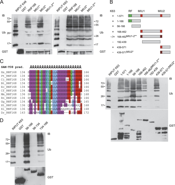
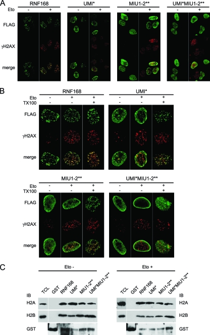
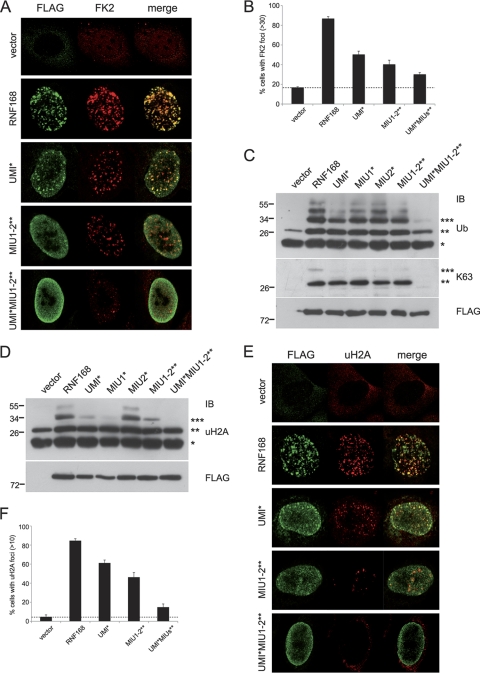
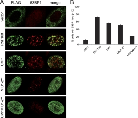
Ubiquitination regulates important cellular processes, including the DNA damage response (DDR) and DNA repair. The complexity of the ubiquitin-mediated signals is decoded by ubiquitin receptors, which contain protein modules named ubiquitin binding domains (UBDs). We previously identified a new ubiquitin ligase, RNF168, involved in DDR and endowed with two UBDs named MIU (motif interacting with ubiquitin). Here we have provided the identification of a novel UBD, the UMI (UIM- and MIU-related UBD), present in RNF168, and characterized the interaction surface with ubiquitin, centered on two Leu residues. We have demonstrated that integrity of the UMI, in addition to the MIUs, is necessary for the proper localization of RNF168 and for ubiquitination of nuclear proteins, including histone H2A. Finally, we have shown that simultaneous inactivation of UMI and MIUs prevents the recruitment to DDR foci of the crucial downstream mediator 53BP1.


Figures

FIG. 1.
FIG. 1.
Identification of a new Ub binding region in RNF168. (A) We performed an in vitro pulldown assay using the indicated GST-tagged RNF168 constructs. GST fusion proteins were incubated with synthetic K48-linked (left panel) or K63-linked (right panel) poly-Ub2-7 chains and separated by SDS-PAGE. Immunoblotting (IB) was performed with antibodies directed against Ub and GST, as described in Materials and Methods. (B) Schematic representation of RNF168 deletion constructs used in pulldown experiments (numbers refer to the amino acid positions within the sequence; RF, RING finger domain); their ability to bind K63 poly-Ub chains, resumed on the left, is shown in the anti-Ub immunoblot of the in vitro pulldown assay (lower panel). Normalization is visualized by anti-GST immunoblotting. (C) Multiple alignments of region 134 to 166 RNF168 homologues in vertebrates. Secondary structure prediction (pred.) was obtained using SAM-T08, a hidden Markov model (HMM)-based protein structure prediction software program (http://compbio.soe.ucsc.edu/SAM_T08/T08-query.html). (D) Mapping of the minimal sequence responsible for Ub binding. An in vitro pulldown assay was performed using the indicated GST-tagged deletion mutants of RNF168, incubated with K63 poly-Ub chains. IB was performed with anti-Ub and anti-GST antibodies.
FIG. 2.
FIG. 2.
Mapping of the amino acid residues required for Ub binding. (A) Comparison of the UMI, UIM, and MIU motifs, where residues involved in the binding to Ub are in bold; underlined sequences were drawn as projected helices using the Helical Wheel Projections software tool (http://rzlab.ucr.edu/scripts/wheel/wheel.cgi). (B) The point mutants were generated by introduction of single (A151G, L149A, or L150A) or double (L149A and L150A) amino acid substitutions addressing the sequence 134 to 166 and tested by in vitro pulldown assay as in Fig. 1D. (C) The indicated RNF168 point mutations were inserted within the sequence encompassing amino acids 56 to 571 and analyzed by in vitro pulldown assay using K48 (left panel) and K63 (right panel) poly-Ub chains. Anti-Ub and anti-GST immunoblots are shown.
FIG. 3.
FIG. 3.
Inactivation of the UMI domain affects localization of RNF168. (A) U2OS cells conditionally expressing RNF168-targeting shRNA were transfected with the indicated FLAG-tagged shRNA-resistant constructs. After 24 h, cells were treated with etoposide for 1 h or left untreated (Eto + and −, respectively). (B) The U2OS cells, treated as described for panel A, were either fixed (TX100 −) or pretreated with Triton X-100 before fixing (TX100 +). Immunostaining of both panels was performed using anti-FLAG and anti-phospho-H2AX (γH2AX) antibodies. (C) A Pulldown assay was performed using GST-RNF168 constructs and chromatin extracted from 293T cells treated or not with etoposide. H2A and H2B antibodies (upper and lower panels, respectively) revealed the presence of specific bands (of about 15 kDa) in all RNF168 constructs but not in the lane with GST alone.
FIG. 4.
FIG. 4.
UMI, MIU1, and MIU2 are required for the ubiquitination events mediated by RNF168. (A) U2OS shRNF168 cells transfected either with the indicated FLAG-tagged shRNA-resistant constructs or with the vector alone were treated with etoposide for 1 h before fixing and immunostained using anti-FLAG and anti-Ub (FK2) antibodies. (B) Quantitation of RNF168-positive cells with more than 30 foci labeled with anti-FK2. (C) Acid extraction of histones from 293T cells transfected with the indicated FLAG-tagged RNF168 constructs. Ubiquitinated proteins were detected by immunoblotting using P4D1 (Ub; upper panel) and Apu3.A8 (K63; medium panel) antibodies. Cell extracts were analyzed for equal expression of the different constructs (FLAG; lower panel). (D) 293T cells were processed as described for panel C and immunodecorated with antibody directed to ubiquitinated forms of histone H2A (uH2A). Cell loading was normalized by FLAG immunoblotting. (E) Immunofluorescence analysis was performed with anti-FLAG and with anti-uH2A antibodies. (F) Quantitation of RNF168-positive cells with more than 10 foci labeled with anti-Ub.
FIG. 5.
FIG. 5.
Inactivation of RNF168 UBDs abolishes 53BP1 recruitment to DDR foci. (A) The U2OS cells conditionally expressing RNF168-targeting shRNA were transfected with the indicated FLAG-tagged shRNA-resistant RNF168 constructs. Twenty-four hours after transfection, cells were treated with etoposide for 1 h, fixed, and immunostained with anti-FLAG and anti-53BP1 antibodies. (B) Quantitation of the two independent experiments described for panel A, representing the percentage of transfected cells with 53BP1-positive foci.

Similar articles

Cited by

References

    1. Acconcia, F., S. Sigismund, and S. Polo. 2009. Ubiquitin in trafficking: the network at work. Exp. Cell Res. 315:1610-1618. - PubMed
    1. Bergink, S., and S. Jentsch. 2009. Principles of ubiquitin and SUMO modifications in DNA repair. Nature 458:461-467. - PubMed
    1. Bhoj, V. G., and Z. J. Chen. 2009. Ubiquitylation in innate and adaptive immunity. Nature 458:430-437. - PubMed
    1. Chen, Z. J., and L. J. Sun. 2009. Nonproteolytic functions of ubiquitin in cell signaling. Mol. Cell 33:275-286. - PubMed
    1. Dikic, I., S. Wakatsuki, and K. J. Walters. 2009. Ubiquitin-binding domains—from structures to functions. Nat. Rev. Mol. Cell Biol. 10:659-671. - PMC - PubMed

Publication types

MeSH terms