. 2011 Jan;79(1):153-66.
doi: 10.1128/IAI.00925-10. Epub 2010 Nov 1.

Role of purine biosynthesis in Bacillus anthracis pathogenesis and virulence

Affiliations

Role of purine biosynthesis in Bacillus anthracis pathogenesis and virulence

Amy Jenkins et al. Infect Immun. 2011 Jan.

Abstract

Bacillus anthracis, the etiological agent of anthrax, is a spore-forming, Gram-positive bacterium and a category A biothreat agent. Screening of a library of transposon-mutagenized B. anthracis spores identified a mutant displaying an altered phenotype that harbored a mutated gene encoding the purine biosynthetic enzyme PurH. PurH is a bifunctional protein that catalyzes the final steps in the biosynthesis of the purine IMP. We constructed and characterized defined purH mutants of the virulent B. anthracis Ames strain. The virulence of the purH mutants was assessed in guinea pigs, mice, and rabbits. The spores of the purH mutants were as virulent as wild-type spores in mouse intranasal and rabbit subcutaneous infection models but were partially attenuated in a mouse intraperitoneal model. In contrast, the purH mutant spores were highly attenuated in guinea pigs regardless of the administration route. The reduced virulence in guinea pigs was not due solely to a germination defect, since both bacilli and toxins were detected in vivo, suggesting that the significant attenuation was associated with a growth defect in vivo. We hypothesize that an intact purine biosynthetic pathway is required for the virulence of B. anthracis in guinea pigs.


Figures

FIG. 1.
FIG. 1.
(A) Purine biosynthetic pathway in B. anthracis as determined by in silico analysis with the KEGG database application. (B) Schematic representation of the gene arrangement of the purine biosynthetic operon in B. anthracis.
FIG. 2.
FIG. 2.
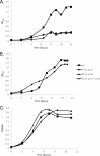
Representative growth curves for the wild-type (wt), purH::Kan, ΔpurH, and ΔpurH + purH strains. The strains were grown in the minimal defined R-medium (A and C) or in R-medium supplemented with 1 mM inosine (B and D). These data represent the results of at least two experiments.
FIG. 3.
FIG. 3.
Reverse transcription-PCR was performed on total RNA harvested from cultures of B. anthracis. The reactions used primers specific for an internal fragment of the purD gene (approximately 380 bp). Lane 1, wild-type RNA; lane 2, RNA collected from the ΔpurH strain; lane 3, RNA collected from the ΔpurD mutant strain; lane 4, RNA collected from the ΔpurH + purH strain. Lane 5, 1 KB Plus ladder; bands shown, from bottom to top, are 200 bp, 300 bp, and 400 bp. Lanes 6 to 9, corresponding negative controls to confirm the absence of DNA from the RNA samples. Additionally, positive controls (not shown) were run using primers specific for the pagA gene as an internal control to ensure the quality of the harvested RNA.
FIG. 4.
FIG. 4.
Representative growth curves for the wild type (wt) and for the ΔpurH, ΔpurD, and ΔpurD + purD mutant strains. The strains were grown in the minimal defined R-medium (A and B) or in R-medium supplemented with 1 mM inosine (C). These data are representative of at least two experiments.
FIG. 5.
FIG. 5.
Mouse intraperitoneal and intranasal challenges with wild-type (WT) Ames or purH mutant spores. (A) BALB/c mice were challenged intraperitoneally with approximately 3 × 10³ wild-type Ames, 3 × 10³ purH::Kan (P, 0.09 for the survival curve and 0.6 for the percentage of mice surviving), or 2.3 × 10³ ΔpurH (P, 0.023 for the survival curve and 0.6 for the percentage of mice surviving) spores. (B) BALB/c mice were challenged intranasally with approximately 3.2 × 10⁶ wild-type Ames, purH::Kan, or ΔpurH spores delivered in 50 μl. These data represent at least two experiments.
FIG. 6.
FIG. 6.
Guinea pig challenges with wild-type (WT) Ames or purH mutant strains. (A) Competitive index generated from the guinea pig assay for comparison of the in vivo fitness of purH mutant strains to that of the wild-type Ames strain (P < 0.0001). Guinea pigs were challenged intramuscularly with approximately 1 × 10³ spores consisting of approximately equal ratios of wild-type and mutant spores. (B) Guinea pigs were challenged intramuscularly with approximately 1 × 10³ wild-type Ames, purH::Kan, or ΔpurH spores (P, <0.0001 for both the percentage of animals surviving and the survival curve), (C) Guinea pigs were challenged intraperitoneally with approximately 4 × 10⁴ wild-type Ames or ΔpurH spores (P, 0.025 for the percentage of animals surviving and 0.04 for the survival curve). (D) Guinea pigs were challenged intraperitoneally with approximately 1.6 × 10⁵ CFU of wild-type Ames or purH::Kan bacilli (P, 0.008 for the percentage of animals surviving and 0.003 for the survival curve).
FIG. 7.
FIG. 7.
LD50 results for ΔpurH spores injected intramuscularly into guinea pigs.
FIG. 8.
FIG. 8.
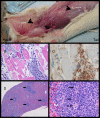
Guinea pigs infected with the ΔpurH strain of B. anthracis. Moribund guinea pigs appeared to be “swollen.” (A) Marked subcutaneous and fascial plane edema. Note the thickened and glistening (marked edema) subcutaneous tissues (arrows). (B) Skeletal muscle of a guinea pig infected with the ΔpurH strain of B. anthracis. There is skeletal muscle degeneration and necrosis characterized by variation in the size and shape of skeletal muscle fibers, loss of cross-striations, and fractured and vacuolated sarcoplasm (cytoplasm). The connective tissue between muscle fibers is markedly expanded by myriad bacilli, polymorphonuclear inflammatory cells, and clear space (edema). HE was used for staining; magnification, ×20. (C) Skeletal muscle, at the injection site, of a guinea pig infected with the ΔpurH strain of B. anthracis. There are myriad strongly positive bacilli within the connective tissue between muscle fibers and invading skeletal muscle fibers. The strong IHC positivity suggests that the bacilli in the muscle produced robust capsules. Immunohistochemistry was carried out for the B. anthracis capsule antigen, with a hematoxylin counterstain; magnification, ×60. (D) Spleen of a guinea pig infected with the ΔpurH strain of B. anthracis. There is diffuse paucity of splenic white pulp. There are scattered normal periarteriolar lymphoid sheaths (arrows). HE was used for staining; magnification, ×4. (E) Spleen of a guinea pig infected with the ΔpurH strain of B. anthracis. Lymphocytolysis (arrows) is evident within the germinal center of a splenic corpuscle. HE was used for staining; magnification, ×40.
FIG. 9.
FIG. 9.
Guinea pig challenges with wild-type (WT) Ames or complemented mutant strains. Guinea pigs were challenged intramuscularly with approximately 1 × 10³ Ames, ΔpurH (P, 0.017 for the percentage of guinea pigs surviving and 0.0240 for the survival curve compared with the WT), ΔpurH + purH (P, 1 for the percentage of guinea pigs surviving, 0.024 for the survival curve, and <0.0001 for TTD compared with the WT), ΔpurH + purD (P, 1 for the percentage of guinea pigs surviving and 0.3 for the survival curve compared with those for the ΔpurH mutant), or ΔpurD + purD (P, ≥0.05 for all tests conducted compared with the WT) spores.

Similar articles

Cited by

References

    1. Aiba, A., and K. Mizobuchi. 1989. Nucleotide sequence analysis of genes purH and purD involved in the de novo purine nucleotide biosynthesis of Escherichia coli. J. Biol. Chem. 264:21239-21246. - PubMed
    1. Allen, S., J. L. Zilles, and D. M. Downs. 2002. Metabolic flux in both the purine mononucleotide and histidine biosynthetic pathways can influence synthesis of the hydroxymethyl pyrimidine moiety of thiamine in Salmonella enterica. J. Bacteriol. 184:6130-6137. - PMC - PubMed
    1. Beaman, T. C., A. D. Hitchins, K. Ochi, N. Vasantha, T. Endo, and E. Freese. 1983. Specificity and control of uptake of purines and other compounds in Bacillus subtilis. J. Bacteriol. 156:1107-1117. - PMC - PubMed
    1. Beedham, R. J., P. C. Turnbull, and E. D. Williamson. 2001. Passive transfer of protection against Bacillus anthracis infection in a murine model. Vaccine 19:4409-4416. - PubMed
    1. Bennett, E. L. 1953. Incorporation of adenine into nucleotides and nucleic acids of C57 mice. Biochim. Biophys. Acta 11:487-496. - PubMed

Publication types