. 2010 Nov 16;107(46):20075-80.
doi: 10.1073/pnas.1013559107. Epub 2010 Nov 1.

Molecular architecture of the vesicular stomatitis virus RNA polymerase

Affiliations

Molecular architecture of the vesicular stomatitis virus RNA polymerase

Amal A Rahmeh et al. Proc Natl Acad Sci U S A. .

Abstract

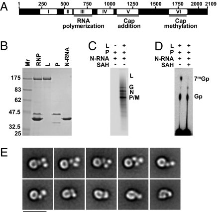
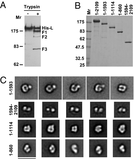
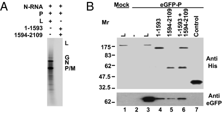
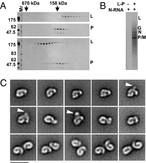
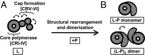
Nonsegmented negative-strand (NNS) RNA viruses initiate infection by delivering into the host cell a highly specialized RNA synthesis machine comprising the genomic RNA completely encapsidated by the viral nucleocapsid protein and associated with the viral polymerase. The catalytic core of this protein-RNA complex is a 250-kDa multifunctional large (L) polymerase protein that contains enzymatic activities for nucleotide polymerization as well as for each step of mRNA cap formation. Working with vesicular stomatitis virus (VSV), a prototype of NNS RNA viruses, we used negative stain electron microscopy (EM) to obtain a molecular view of L, alone and in complex with the viral phosphoprotein (P) cofactor. EM analysis, combined with proteolytic digestion and deletion mapping, revealed the organization of L into a ring domain containing the RNA polymerase and an appendage of three globular domains containing the cap-forming activities. The capping enzyme maps to a globular domain, which is juxtaposed to the ring, and the cap methyltransferase maps to a more distal and flexibly connected globule. Upon P binding, L undergoes a significant rearrangement that may reflect an optimal positioning of its functional domains for transcription. The structural map of L provides new insights into the interrelationship of its various domains, and their rearrangement on P binding that is likely important for RNA synthesis. Because the arrangement of conserved regions involved in catalysis is homologous, the structural insights obtained for VSV L likely extend to all NNS RNA viruses.


Conflict of interest statement

The authors declare no conflict of interest.

Figures

Fig. 1.
Fig. 1.
Functional and structural organization of VSV L. (A) A schematic of the linear map of VSV L depicts the six conserved regions (CR I–VI) among NNS virus L proteins as white boxes separated by variable regions shown in black. The RNA polymerization, cap addition, and cap methylation activities have been mapped to CRIII, CRV, and CRVI, respectively. (B) Purification of recombinant RNP components. L and P were individually expressed and purified, and N–RNA was purified from recombinant VSV as previously described (4). Proteins were analyzed by 10% SDS/PAGE and stained with Coomassie blue, Mr, molecular weight marker. (C) Reconstitution of RNA synthesis from purified N–RNA, P, and L. Transcription reactions were performed in the presence of [α-32P] GTP and the products analyzed by electrophoresis on acid-agarose gels. The identity of the five VSV mRNAs P, M (matrix), N, G (glycoprotein), and L is shown at Right. (D) Analysis of the mRNA cap structure. Transcription reactions were performed as in C in the absence or presence of the MTase inhibitor S-adenosyl homocysteine (SAH). The RNA products were digested with TAP and the products resolved by TLC. The mobilities of Gp and 7mGp are indicated at Right. (E) EM characterization of L. The 10 presented class averages show the ring-shaped core domain and illustrate the structural variability of the appendage. (Scale bar: 20 nm.)
Fig. 2.
Fig. 2.
The structural domains of VSV L. (A) Limited proteolysis of L. Purified L was digested with trypsin, before separation by 6% SDS/PAGE. The cleavage products were detected using an anti-His antibody that recognizes the N-terminal tag. Fragments with intact N termini that were selected for further analysis are identified as F1, F2, and F3. (B) Fragments corresponding to those released by trypsin digestion (1–1,593 for F1, 1–1,114 for F2, and 1–860 for F3) and a complementary fragment to F1 (1,594–2,109) were designed with an N-terminal 6× His tag, expressed and purified. The purified proteins were separated by 10% SDS/PAGE and visualized by Coomassie blue. (C) Electron microscopic characterization of the L fragments. The class averages show the structural features of the various L fragments. (Scale bar: 20 nm.)
Fig. 3.
Fig. 3.
Functional analysis of the complementary N- and C-terminal fragments of VSV L. (A) RNA synthesis. Equimolar quantities of L or 1–1,593 + 1,594–2,109 were used to reconstitute RNA synthesis in vitro and the products analyzed as in Fig 1C. (B) P protein binding. Full-length L, or fragments 1–1,593, 1,594–2,109, 1–1,593 + 1,594–2,109 were used as bait to pull down eGFP–P expressed in BSRT7 cells. Western blots show the His-tagged proteins (Upper) and the corresponding captured eGFP–P (Lower). Mock transfected BSRT7 cells (lane 1), no L (lane 2), and His-tagged N-terminal domain of anthrax lethal factor (lane 7) are shown as specificity controls.
Fig. 4.
Fig. 4.
Functional and structural characterization of the L–P complex. (A) Size exclusion chromatography of L (Top), P (Middle), and L–P (Bottom). The fractions eluted from a Superdex 200 column were analyzed by 10% SDS/PAGE and the proteins visualized by Coomassie blue. The elution positions of two Mwt standards are indicated by arrows. (B) RNA synthesis in vitro. Transcription reactions were reconstituted using N–RNA and the L–P complex peak fraction and the products analyzed as in Fig 1C. (C) EM characterization of L–P. The Top two rows show representative averages of the single L–P complexes and the Bottom row shows representative averages of the double species. The arrowheads indicate the additional globular density occasionally seen in averages of the single L–P complexes. (Scale bar: 20 nm.)
Fig. 5.
Fig. 5.
Molecular architecture of VSV L. (A) L is organized into a core ring structure harboring the RdRP domain and a flexible appendage containing the activities necessary for cap formation (capping + methylation). The arrows depict putative flexible linkers. (B) Upon binding to P, L undergoes a structural rearrangement. The L–P complex exists as a monomer or a dimer in which the L pairs are likely bridged by interaction with an oligomer of P. The arrow represents the variable orientation of L proteins in the dimers.

References

    1. Emerson SU, Wagner RR. Dissociation and reconstitution of the transcriptase and template activities of vesicular stomatitis B and T virions. J Virol. 1972;10:297–309. - PMC - PubMed
    1. Li J, Fontaine-Rodriguez EC, Whelan SP. Amino acid residues within conserved domain VI of the vesicular stomatitis virus large polymerase protein essential for mRNA cap methyltransferase activity. J Virol. 2005;79:13373–13384. - PMC - PubMed
    1. Sleat DE, Banerjee AK. Transcriptional activity and mutational analysis of recombinant vesicular stomatitis virus RNA polymerase. J Virol. 1993;67:1334–1339. - PMC - PubMed
    1. Li J, Rahmeh A, Morelli M, Whelan SP. A conserved motif in region V of the large polymerase proteins of nonsegmented negative-sense RNA viruses that is essential for mRNA capping. J Virol. 2008;82:775–784. - PMC - PubMed
    1. Barr JN, Whelan SP, Wertz GW. cis-Acting signals involved in termination of vesicular stomatitis virus mRNA synthesis include the conserved AUAC and the U7 signal for polyadenylation. J Virol. 1997;71:8718–8725. - PMC - PubMed

Publication types

MeSH terms

LinkOut - more resources