. 2010 Nov 16;107(46):19927-32.
doi: 10.1073/pnas.1007381107. Epub 2010 Nov 1.

Nicotinic acid adenine dinucleotide phosphate regulates skeletal muscle differentiation via action at two-pore channels

Affiliations

Nicotinic acid adenine dinucleotide phosphate regulates skeletal muscle differentiation via action at two-pore channels

Parvinder K Aley et al. Proc Natl Acad Sci U S A. .

Erratum in

  • Proc Natl Acad Sci U S A. 2011 Mar 22;108(12):5139

Abstract

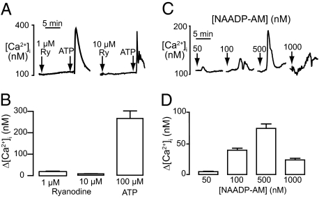
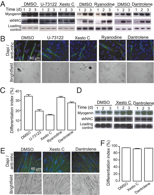
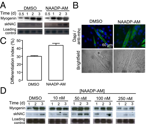
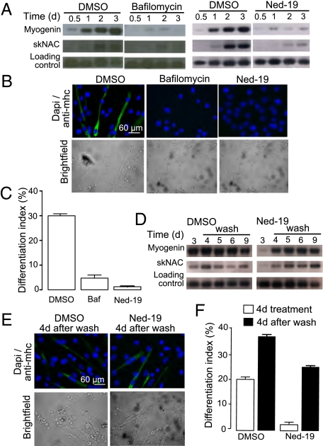
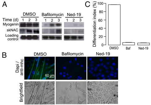
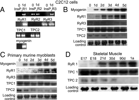
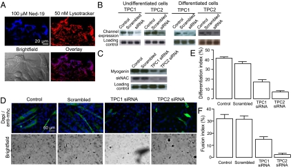
Calcium signaling is essential for the differentiation of many cell types, including skeletal muscle cells, but its mechanisms remain elusive. Here we demonstrate a crucial role for nicotinic acid adenine dinucleotide phosphate (NAADP) signaling in skeletal muscle differentiation. Although the inositol trisphosphate pathway may have a partial role to play in this process, the ryanodine signaling cascade is not involved. In both skeletal muscle precursors and C2C12, cells interfering with NAADP signaling prevented differentiation, whereas promoting NAADP signaling potentiated differentiation. Moreover, siRNA knockdown of two-pore channels, the target of NAADP, attenuated differentiation. The data presented here strongly suggest that in myoblasts, NAADP acts at acidic organelles on the recently discovered two-pore channels to promote differentiation.


Conflict of interest statement

The authors declare no conflict of interest.

Figures

Fig. 1.
Fig. 1.
NAADP induces calcium release in undifferentiated C2C12 cells. (A) Example traces of calcium release induced by 1 or 10 μM ryanodine (first arrow) followed by 100 μM ATP (second arrow). (B) Bar graph (mean with SEM; n = 25–40 cells) representing the change in cytosolic calcium following addition of ryanodine or ATP. (C) Example traces of calcium release induced by 50, 100, 500, and 1 μM NAADP-AM. Arrows indicate addition of NAADP-AM. (D) Bar graph (mean with SEM; n = 12–16 cells) representing the change in cytosolic calcium following addition of NAADP-AM.
Fig. 2.
Fig. 2.
Neither the InsP3 or ryanodine signaling pathway is essential for skeletal muscle differentiation. C2C12 cells were induced to differentiate in the presence of 1 μM xestospongin C, 5 μM U-73122, 100 μM ryanodine, or 100 μM dantrolene. (A) RNA was harvested at the indicated time points and analyzed for expression of myogenin and skNAC by Northern blot analysis. (B) Following 4 d of differentiation, cells were stained for myosin heavy chain and DAPI to determine the differentiation index (percent nuclei in myosin heavy chain-positive cells). (C) Bar chart (mean with SEM; n = 4) representing the differentiation index following treatment for 4 d with DMSO, or an inhibitor of the IP3 or ryanodine signaling pathway. Primary murine myoblast were induced to differentiate in the presence of 1 μM xestospongin C, 5 μM U-73122, 100 μM dantrolene, or 100 μM ryanodine. (D) RNA was harvested at the indicated time points and analyzed for expression of myogenin and skNAC by Northern blot analysis. (E) Following 4 d of differentiation, cells were stained for myosin heavy chain and DAPI to determine the differentiation index (percent nuclei in myosin heavy chain-positive cells). (F) Bar chart (mean with SEM; n = 4) representing the differentiation index following treatment for 4 d with DMSO, or an inhibitor of the IP3 or ryanodine signaling pathway.
Fig. 3.
Fig. 3.
NAADP-AM promotes skeletal muscle differentiation. C2C12 cells were induced to differentiate in the presence or absence of 50 nM NAADP-AM. (A) RNA was harvested at the indicated time points and analyzed for expression of myogenin and skNAC by Northern blot using specific probes. (B) Following 4 d of differentiation, cells were stained for myosin heavy chain and DAPI to determine the differentiation index (percent nuclei in myosin heavy chain-positive cells). (C) Bar chart (mean with SEM; n = 4) representing the differentiation index following treatment for 4 d with either DMSO or NAADP-AM. (D) Primary murine myoblast were induced to differentiate in the presence or absence of 10, 50, 100, or 250 nM NAADP-AM. RNA was harvested at the indicated time points and analyzed for the expression of myogenin and skNAC by Northern blot analysis.
Fig. 4.
Fig. 4.
NAADP signaling is essential for differentiation of C2C12 cells. C2C12 cells were induced to differentiate in the presence of DMSO, 200 nM bafilomycin, or 100 μM Ned-19. (A) RNA was harvested at the indicated time points and analyzed for expression of myogenin and skNAC by Northern blot analysis. (B) Following 4 d of differentiation, cells were stained for myosin heavy chain and DAPI to determine the differentiation index (percent nuclei in myosin heavy chain-positive cells). (C) Bar chart (mean with SEM; n = 4) representing the differentiation index following treatment for 4 d with control, bafilomycin, or Ned-19. Recovery of C2C12 differentiation following removal of Ned-19 was demonstrated by (D) Northern blot, (E) myosin heavy chain and DAPI staining, and (F) calculation of the differentiation index.
Fig. 5.
Fig. 5.
NAADP signaling is essential for differentiation of primary murine myoblasts. Primary murine myoblast were differentiated in the presence of with 200 nM bafilomycin or 100 μM Ned-19. (A) RNA was harvested at the indicated time points and analyzed for the expression of myogenin and skNAC by Northern blot analysis. (B) Following 4 d of differentiation, cells were stained for myosin heavy chain and DAPI to determine the differentiation index (percent nuclei in myosin heavy chain-positive cells). (C) Bar chart (mean with SEM; n = 4) representing the differentiation index following treatment for 4 d with control, bafilomycin, or Ned-19.
Fig. 6.
Fig. 6.
Calcium channel expression during skeletal muscle differentiation. C2C12 cells were induced to differentiate. (A) Before and after 24 h of differentiation the RNA was harvested and analyzed for expression of InsP3R, RyR, and TPC subtypes by RT-PCR with specific primers. Differentiation was monitored by detection of myogenin. (B) RNA was harvested at the indicated time points and analyzed using Northern blot for expression of RyR and TPC subtypes, using specific probes. Differentiation was monitored by detection of myogenin. (C) Primary murine myoblasts were induced to differentiate. RNA was harvested at the indicated time points and analyzed for the expression of RyR and TPC subtypes, using specific probes. Differentiation was monitored by detection of myogenin. (D) RNA from mouse skeletal muscle at different stages of development was analyzed using Northern blot for the expression of TPC1, TPC2, and RyR1 using specific probes.
Fig. 7.
Fig. 7.
C2C12 differentiation is altered by down-regulation of TPCs on acidic organelles. (A) Confocal microscopy of undifferentiated C2C12 cells. (Upper Left) Labeling of NAADP receptor with 100 μM Ned-19. (Upper Right) Labeling of acidic organelles with 50 nM lysotracker. (Lower Left) Bright field image of C2C12 cells. (Lower Right) Overlay of Ned-19 and lysotracker labeling showing colocalization. (B) C2C12 cells were transfected with siRNA against TPC1 or TPC2, respectively. RNA was harvested either 24 h after transfection (undifferentiated cells) or 24 h after differentiation was induced (differentiated cells). The RNA was analyzed for expression of TPC1 or TPC2, respectively, by Northern blot analysis. (C) C2C12 cells were transfected with siRNA against TPC1 or TPC2 for 24 h and differentiated for 24 h. RNA was harvested and analyzed for expression of myogenin and skNAC by Northern blot analysis. (D) C2C12 cells were transfected with siRNA against TPC1 or TPC2 for 24 h and differentiated for 4 d. The cells were stained for myosin heavy chain and DAPI to determine the differentiation index (percent nuclei in myosin heavy chain-positive cells). (E) Bar chart (mean with SEM; n = 4) representing the differentiation index following treatment for 4 d with control, scrambled siRNA, TPC1 siRNA, or TPC2 siRNA. (F) Bar chart (mean with SEM; n = 4) representing the fusion index (percent nuclei in myosin heavy chain-positive cells with at least three nuclei) following treatment for 4 d with control, scrambled siRNA, TPC1 siRNA, or TPC2 siRNA.

References

    1. Berridge MJ, Bootman MD, Roderick HL. Calcium signalling: Dynamics, homeostasis and remodelling. Nat Rev Mol Cell Biol. 2003;4:517–529. - PubMed
    1. Ikura M, Osawa M, Ames JB. The role of calcium-binding proteins in the control of transcription: structure to function. Bioessays. 2002;24:625–636. - PubMed
    1. Hogan PG, Chen L, Nardone J, Rao A. Transcriptional regulation by calcium, calcineurin, and NFAT. Genes Dev. 2003;17:2205–2232. - PubMed
    1. Galione A, Ruas M. NAADP receptors. Cell Calcium. 2005;38:273–280. - PubMed
    1. Dammermann W, et al. NAADP-mediated Ca2+ signaling via type 1 ryanodine receptor in T cells revealed by a synthetic NAADP antagonist. Proc Natl Acad Sci USA. 2009;106:10678–10683. - PMC - PubMed

Publication types

LinkOut - more resources