. 2010 Nov 16;107(46):19903-8.
doi: 10.1073/pnas.1007404107. Epub 2010 Nov 1.

Tumor formation initiated by nondividing epidermal cells via an inflammatory infiltrate

Affiliations

Tumor formation initiated by nondividing epidermal cells via an inflammatory infiltrate

Esther N Arwert et al. Proc Natl Acad Sci U S A. .

Abstract

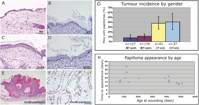
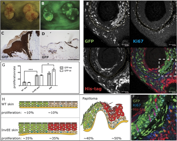
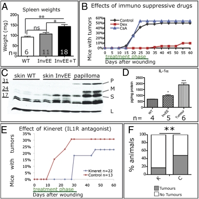
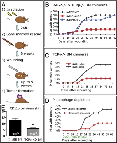
In mammalian epidermis, integrin expression is normally confined to the basal proliferative layer that contains stem cells. However, in epidermal hyperproliferative disorders and tumors, integrins are also expressed by suprabasal cells, with concomitant up-regulation of Erk mitogen-activated protein kinase (MAPK) signaling. In transgenic mice, expression of activated MAPK kinase 1 (MEK1) in the suprabasal, nondividing, differentiated cell layers (InvEE transgenics) results in epidermal hyperproliferation and skin inflammation. We now demonstrate that wounding induces benign tumors (papillomas and keratoacanthomas) in InvEE mice. By generating chimeras between InvEE mice and mice that lack the MEK1 transgene, we demonstrate that differentiating, nondividing cells that express MEK1 stimulate adjacent transgene-negative cells to divide and become incorporated into the tumor mass. Dexamethasone treatment inhibits tumor formation, suggesting that inflammation is involved. InvEE skin and tumors express high levels of IL1α; treatment with an IL1 receptor antagonist delays tumor onset and reduces incidence. Depletion of γδ T cells and macrophages also reduces tumor incidence. Because a hallmark of cancer is uncontrolled proliferation, it is widely assumed that tumors arise only from dividing cells. In contrast, our studies show that differentiated epidermal cells can initiate tumor formation without reacquiring the ability to divide and that they do so by triggering an inflammatory infiltrate.


Conflict of interest statement

The authors declare no conflict of interest.

Figures

Fig. 1.
Fig. 1.
Wound-induced papillomas in InvEE transgenic mice. (A, C, and E) H&E- and (B, D, and F) Ki67-stained sections of nontransgenic (WT: A and B) and InvEE transgenic (C and D) back skin and InvEE wound-induced papilloma (E and F). (Scale bar: 100 μm.) (G) Incidence of spontaneous (“spont”: purple and mauve) and wound-induced (“wnd”: yellow and blue) papillomas in male (M) and female (F) InvEE transgenic mice. Error bars: 95% confidence intervals. (H) Time between wounding and appearance of papillomas in InvEE mice of different ages.
Fig. 2.
Fig. 2.
Papilloma formation in GFP/InvEE chimeric mice. (A and B) Macroscopic appearance of two wound-induced papillomas, viewed under natural (A) and fluorescent (B) light. (C and D) GFP immunohistochemistry of papilloma (C) and skin (D) from GFP/InvEE mice. (E and F) GFP/InvEE papilloma labeled with antibodies to GFP (green), Ki67 (blue), and MEK1 transgene His tag (red). Each label is shown separately in black and white; merge is in color. Dashed lines in E demarcate basement membrane. (F) Enlarged view of boxed area in E, with GFP-negative, Ki67-positive basal cells indicated with orange arrows and GFP-positive, Ki67-positive cells indicated with pink arrows. [Scale bars: 200 μm (C and D); 50 μm (E).] (G) Percentage of GFP-positive (GFP+ve) or -negative (GFP-ve) basal cells that were Ki67 positive in GFP/WT skin (WT; n = 3), GFP/InvEE skin (InvEE skin; n = 4), and GFP/InvEE papillomas (“pap”: n = 4). *P < 0.05, ***P < 0.001. (H) Data in G shown schematically to illustrate relationship of GFP-positive (green) and -negative (beige) basal cells to the overlying suprabasal cells (GFP positive: green; GFP negative, MEK1 transgene positive: red).
Fig. 3.
Fig. 3.
Effect of IL1 on wound-induced tumor formation. (A) Spleen weights (mg) of wild type (WT), InvEE mice, and tumor-bearing InvEE mice (InvEE+T) with number of mice indicated. ns: not significant. (B) Effects of cyclosporine A and dexamethasone on wound-induced tumor formation. (C) Western blot of wild-type (WT) skin, InvEE skin, and InvEE papilloma lysates. Each track is from a different mouse. Blot probed with anti-IL1α or, as a loading control (L), β-tubulin. P: pro-IL1α; M: membrane-bound IL1α; S: soluble IL1α. (D) Quantitation of IL1α in skin and tumor protein lysates with cytometric beads. (E and F) Effect of Kineret on wound-induced tumor formation (E) and tumor incidence (F). F: n = 36 Kineret-treated (K), n = 29 controls (C). *P < 0.05, **P < 0.01, ***P < 0.001.
Fig. 4.
Fig. 4.
Effect of bone marrow-derived cells on wound-induced tumor formation. (A) Schematic of bone marrow transplantation protocol. (B and C) Effect on wound-induced papilloma formation of reconstitution with Rag2 −/− (B), TCRβ −/− (B), and TCRδ−/− (C) bone marrow. (D) Effect of macrophage depletion with clodronate liposomes. (E) Number of cells expressing CD11bhigh per millimeter of dermis in skin of InvEE mice transplanted with InvEE (n = 5) or TCRδ−/− (n = 3) bone marrow (BM). Unpaired t test: t = 3.697, P = 0.0049. **P < 0.01.

References

    1. Janes SM, Watt FM. New roles for integrins in squamous-cell carcinoma. Nat Rev Cancer. 2006;6:175–183. - PubMed
    1. Watt FM. Role of integrins in regulating epidermal adhesion, growth and differentiation. EMBO J. 2002;21:3919–3926. - PMC - PubMed
    1. Yang X, Pursell B, Lu S, Chang TK, Mercurio AM. Regulation of β4-integrin expression by epigenetic modifications in the mammary gland and during the epithelial-to-mesenchymal transition. J Cell Sci. 2009;122:2473–2480. - PMC - PubMed
    1. Haase I, Hobbs RM, Romero MR, Broad S, Watt FM. A role for mitogen-activated protein kinase activation by integrins in the pathogenesis of psoriasis. J Clin Invest. 2001;108:527–536. - PMC - PubMed
    1. Hobbs RM, Silva-Vargas V, Groves R, Watt FM. Expression of activated MEK1 in differentiating epidermal cells is sufficient to generate hyperproliferative and inflammatory skin lesions. J Invest Dermatol. 2004;123:503–515. - PubMed

Publication types

MeSH terms

Substances

LinkOut - more resources