. 2010 Nov 16;107(46):20039-44.
doi: 10.1073/pnas.1014509107. Epub 2010 Nov 1.

Experimental human cytomegalovirus latency in CD14+ monocytes

Affiliations

Experimental human cytomegalovirus latency in CD14+ monocytes

Danna Hargett et al. Proc Natl Acad Sci U S A. .

Abstract

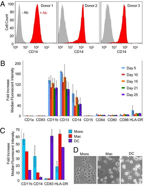
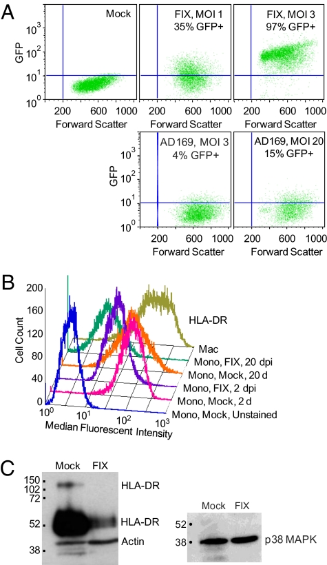
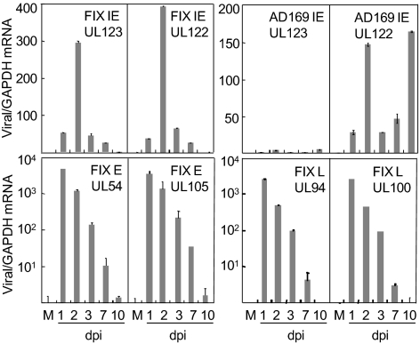
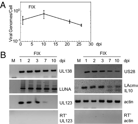
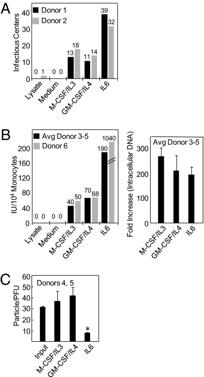
CD14(+) monocytes are a reservoir for latent human cytomegalovirus, and virus replication is reactivated during their differentiation to macrophages or dendritic cells. It has not been clear whether the virus can establish latency upon direct infection of monocytes or whether it must first become quiescent in a progenitor cell that subsequently differentiates to generate a monocyte. We report that infection of primary human monocytes with a clinical strain of human cytomegalovirus exhibits the hallmarks of latency. We established conditions for culturing monocytes that prevent differentiation for at least 25 d, as evidenced by cell surface marker expression. Infection of these monocytes with the FIX clinical strain resulted in transient accumulation of many viral lytic RNAs and sustained expression of four previously described latency-associated transcripts. The amount of viral DNA remained constant after infection, and cell surface and total HLA-DR proteins were substantially reduced on a continuing basis after infection. When treated with cytokine mixtures that stimulate differentiation to a macrophage or dendritic cell phenotype, infected monocytes reactivated virus replication and produced infectious progeny. Treatment of infected monocytes with IL-6 alone also was sufficient for reactivation, and the particles produced after exposure to this cytokine were about fivefold more infectious than virions produced by other treatments. We propose that in vivo microenvironments influence not only the efficiency of reactivation but also the infectivity of the virions produced from latently infected monocytes.


Conflict of interest statement

The authors declare no conflict of interest.

Figures

Fig. 1.
Fig. 1.
Monocyte culture. (A) Reproducible isolation of CD14+ populations. Monocytes from three donors were prepared, maintained in culture for 24 h, and then analyzed for CD14 expression by flow cytometry. Samples receiving no antibody (gray) were compared with samples treated with fluorescently conjugated anti-CD14 antibody (red). (B) Stable expression of cell surface markers over an extended period in culture. Cells from two donors were monitored by flow cytometry with phycoerythrin (PE)-CY7–conjugated anti-CD14 and PE-conjugated antibodies to various marker proteins. Fold increases in median fluorescent intensity relative to isotype controls were calculated for cells from the population of CD14high backgated cells. The mean plus SE are presented. (C) Induction of macrophage and dendritic cell markers in response to cytokine treatment. After 24 d in culture, monocytes (Mono; blue bars) were transferred to standard tissue culture plates in medium containing either M-CSF plus IL-3 (Mac; red bars) or GM-CSF plus IL-4 (DC; purple bars); 6 d later, cell surface markers were analyzed by flow cytometry, and the mean fold increase in median fluorescent intensity (±SE) relative to isotype controls for cells from two donors is displayed. (D) Morphology of undifferentiated vs. differentiated monocytes. Cells were visualized by phase microscopy.
Fig. 2.
Fig. 2.
Infection of monocytes. (A) Cultured monocytes are efficiently infected with the FIX strain of HCMV. After 1 d in culture, monocytes were mock-infected or infected with FIX- or AD169-expressing GFP at the indicated multiplicities of infection (MOI). Two days later, cells were analyzed by flow cytometry for GFP expression. (B) HLA-DR surface expression is reduced after infection of monocytes. Cells were mock-infected or infected with a FIX derivative expressing an IE2–GFP fusion protein and assayed for HLA-DR by flow cytometry at 2 or 20 d post infection (dpi). Cells differentiated to a macrophage (Mac) phenotype were also analyzed as a control. (C) Reduced total cell HLA-DR after infection of monocytes. Cells were mock-infected or infected with FIX and assayed for HLA-DR 5 d later by Western blot. Intact αβ heterodimers and monomers are labeled. The blot was probed for β-actin and p38 MAPK as controls.
Fig. 3.
Fig. 3.
Transient expression of HCMV lytic RNAs in monocytes. One day after isolation, monocytes were mock-infected (M) or infected with FIX or AD169, and RNA was prepared on various days postinfection. Representative immediate-early (IE), early (E), and late (L) viral RNAs were quantified by using RT-qPCR. Samples were assayed in quadruplicate; standard curves were generated using purified viral DNA with a β-actin insertion, and quantities of viral RNAs were normalized to cellular GAPDH RNA levels.
Fig. 4.
Fig. 4.
Genome maintenance and expression of latency-associated RNAs in monocytes. (A) Maintenance of the FIX genome. DNA was isolated from monocytes at various times after infection, and viral DNA was quantified by qPCR. Samples were run in quadruplicate, and viral genomes per cell were calculated by dividing the number of genomes (normalized to actin) by the cell number. ANOVA was performed to test for variation in DNA content over time and yielded a P value of 0.44, supporting the interpretation that the means do not differ. (B) Latency-associated RNAs are present in FIX-infected monocytes throughout a 10-d time course. Total RNA was isolated from monocytes and assayed by RT-PCR (40 cycles) followed by gel electrophoresis of the products. Control assays were performed without reverse transcriptase (RT) to monitor for DNA contamination. Negative images are displayed to better visualize DNA bands.
Fig. 5.
Fig. 5.
Reactivation of FIX by differentiation of monocytes. Ten days before initiating a reactivation assay, CD14+ monocytes were infected with FIX virus (donors 1, 2, and 6) or a derivative of FIX expressing an IE2-GFP fusion protein (donors 3–5). (A) Production of virus by coculture of monocytes with fibroblasts in medium supplemented with cytokines. After 11 d, infectious centers containing more than or equal to five infected cells were counted. As controls, lysates of infected monocytes and medium from infected cultures, prepared at the start of the reactivation experiment, were added to fibroblasts. (B Left) Production of virus by transferring monocytes to standard cell culture plates in medium supplemented with cytokines. After 11 d, supernatants were harvested and assayed for infectious units (IUs; i.e., the number of IE1-positive fibroblasts generated at 24 h postinfection). Monocyte lysates and medium were assayed as controls. (Right) Amplification of viral DNA on differentiation of monocytes. Total cell DNA was prepared (donors 3–5), quantified by qPCR, and reported as the fold increase over the amount of viral DNA in infected monocytes immediately before reactivation. (C) IL-6–mediated reactivation generates virions with enhanced infectivity. Particle to PFU ratios were calculated by dividing the amount of DNA in DNase before samples (Fig. S3) by the number of infectious units for donors 4 and 5. The asterisk indicates a P value < 0.0001 compared with the input virus (produced in fibroblasts) and IL-6–induced virus (produced in differentiating monocytes) by the Student t test.

References

    1. Mocarski ES, Shenk T, Pass RF. Cytomegaloviruses. In: Knipe DM, Howley PM, editors. Fields Virology. 5th Ed. Philadelphia: Wolters Kluwer Health/Lippincott Williams & Wilkins; 2007. pp. 2701–2772.
    1. Sinclair J. Human cytomegalovirus: Latency and reactivation in the myeloid lineage. J Clin Virol. 2008;41:180–185. - PubMed
    1. Goodrum F, Reeves M, Sinclair J, High K, Shenk T. Human cytomegalovirus sequences expressed in latently infected individuals promote a latent infection in vitro. Blood. 2007;110:937–945. - PMC - PubMed
    1. Petrucelli A, Rak M, Grainger L, Goodrum F. Characterization of a novel Golgi apparatus-localized latency determinant encoded by human cytomegalovirus. J Virol. 2009;83:5615–5629. - PMC - PubMed
    1. Schrier RD, Nelson JA, Oldstone MB. Detection of human cytomegalovirus in peripheral blood lymphocytes in a natural infection. Science. 1985;230:1048–1051. - PubMed

Publication types

MeSH terms