. 2010 Nov 16;107(46):19967-72.
doi: 10.1073/pnas.1014669107. Epub 2010 Nov 1.

MyD88 signaling in nonhematopoietic cells protects mice against induced colitis by regulating specific EGF receptor ligands

Affiliations

MyD88 signaling in nonhematopoietic cells protects mice against induced colitis by regulating specific EGF receptor ligands

Katharina Brandl et al. Proc Natl Acad Sci U S A. .

Abstract

Toll-like receptors (TLRs) trigger intestinal inflammation when the epithelial barrier is breached by physical trauma or pathogenic microbes. Although it has been shown that TLR-mediated signals are ultimately protective in models of acute intestinal inflammation [such as dextran sulfate sodium (DSS)-induced colitis], it is less clear which cells mediate protection. Here we demonstrate that TLR signaling in the nonhematopoietic compartment confers protection in acute DSS-induced colitis. Epithelial cells of MyD88/Trif-deficient mice express diminished levels of the epidermal growth factor receptor (EGFR) ligands amphiregulin (AREG) and epiregulin (EREG), and systemic lipopolysaccharide administration induces their expression in the colon. N-ethyl-N-nitrosourea (ENU)-induced mutations in Adam17 (which is required for AREG and EREG processing) and in Egfr both produce a strong DSS colitis phenotype, and the Adam17 mutation exerts its deleterious effect in the nonhematopoietic compartment. The effect of abrogation of TLR signaling is mitigated by systemic administration of AREG. A TLR→MyD88→AREG/EREG→EGFR signaling pathway is represented in nonhematopoietic cells of the intestinal tract, responds to microbial stimuli once barriers are breached, and mediates protection against DSS-induced colitis.


Conflict of interest statement

The authors declare no conflict of interest.

Figures
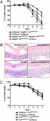
Fig. 1.
Fig. 1.
MyD88 signals in nonhematopoietic cells facilitate susceptibility to acute DSS colitis. (A) Bone marrow chimeras were generated and percent initial weight was determined for 7 d after 2% DSS administration (n = 5 for Myd88−/−/TrifLps2/Lps2Myd88−/−/TrifLps2/Lps2 and n = 8–10 for all other groups; ***P ≤ 0.001 for Myd88−/−/TrifLps2/Lps2→C57BL/6J versus C57BL/6J→Myd88−/−/TrifLps2/Lps2, Mann–Whitney test). (B) Representative photomicrographs (magnification 200×; H&E staining) of colons from different chimeric mice 7 d after 2% DSS administration. (C) Percentage of initial weight from different TrifLps2/Lps2 BM chimeras after 2% DSS administration for 7 d (n = 3 for TrifLps2/Lps2TrifLps2/Lps2 and n = 5 for all other groups).
Fig. 2.
Fig. 2.
TLR-mediated signals are important for Areg and Ereg induction. (A) Gene expression analysis of epithelial cells from C57BL/6J and Myd88−/−/TrifLps2/Lps2 double-deficient mice. The heatmap shows the mean-scaled expression for differently regulated transcripts. (B) mRNA from epithelial cells of Myd88−/−/TrifLps2/Lps2 double-deficient mice were analyzed for the expression of different EGFR ligands. Expression levels were normalized to β-actin. Values shown are expressed relative to the expression of Myd88−/−/TrifLps2/Lps2 double-deficient epithelial cells (n = 5–6; *P ≤ 0.05, **P ≤ 0.01, Mann–Whitney test). (C) Percentage of initial weight from C57BL/6J and Myd88−/−/TrifLps2/Lps2 double-deficient mice treated with either AREG or PBS daily. All mice received 2% DSS in drinking water for 6 d. P values were calculated for Myd88−/−/TrifLps2/Lps2 double-deficient mice treated with AREG versus PBS (n = 4–5; *P ≤ 0.05, Mann–Whitney test). (D) mRNA expression of different EGFR ligands in C57BL/6J epithelial cells before or 2 h after LPS administration (i.p.). Expression levels were normalized to β-actin or 18s. Values shown are expressed relative to the expression in C57BL/6J epithelial cells before LPS administration (n = 5–6; *P ≤ 0.05, Mann–Whitney test). (E) mRNA was extracted from the colon of wild-type mice and mice receiving antibiotics (MNV) for 1, 3, and 7 d. Different EGFR ligand expression was examined by quantitative real-time PCR. Expression levels were normalized to β-actin. Values shown are expressed relative to the expression of wild-type mice receiving no treatment (n = 6–14 mice per group; *P ≤ 0.05, **P ≤ 0.01, each compared with untreated, Mann–Whitney test).
Fig. 3.
Fig. 3.
Mice with defects in EGFR signaling are highly susceptible to DSS-induced colitis. (A) Body weight of C57BL/6J and Egfr+/Velvet mice after the onset of treatment with DSS. *P ≤ 0.05, **P ≤ 0.01, ***P ≤ 0.001, Mann-Whitney test, n = 6–10. (B) The wavedX mutation corresponds to a T→A transversion at position 1186 of the Adam17 transcript. (C) Macrophages were stimulated with LPS, and surface (Left) or intracellular (Middle) TNF was measured by flow cytometry. TNF in the supernatant (Right) was determined by ELISA after 4-h incubation with different LPS concentrations (*P ≤ 0.05, Mann–Whitney test, n = 4 for Adam17+/wavedX and n = 5 for Adam17wavedX/wavedX). Error bars denote SD. MFI, mean fluorescence intensity. (D) Percent initial weight of Adam17+/wavedX mice and Adam17wavedX/wavedX mice (n = 10 for Adam17+/wavedX and n = 4 for Adam17wavedX/wavedX) after 2% DSS administration. **P ≤ 0.01, Mann-Whitney test. (E) Adam17wavedX/wavedX BM chimeras were generated and percent of initial weight was determined after the onset of treatment with DSS for 8 d (n = 9–12 each group; *P ≤ 0.05, Mann–Whitney test). (F) Representative photomicrographs (magnification 200×; H&E staining) of colons from different chimeric mice 7 d after 2% DSS administration. (G) Model of a TLR→MyD88→EGFR ligand→EGFR signaling pathway in nonhematopoietic cells of the intestinal tract.

References

    1. Podolsky DK. Inflammatory bowel disease. N Engl J Med. 2002;347:417–429. - PubMed
    1. Xavier RJ, Podolsky DK. Unravelling the pathogenesis of inflammatory bowel disease. Nature. 2007;448:427–434. - PubMed
    1. Yamamoto-Furusho JK, Podolsky DK. Innate immunity in inflammatory bowel disease. World J Gastroenterol. 2007;13:5577–5580. - PMC - PubMed
    1. Sartor RB. Mechanisms of disease: Pathogenesis of Crohn's disease and ulcerative colitis. Nat Clin Pract Gastroenterol Hepatol. 2006;3:390–407. - PubMed
    1. Rakoff-Nahoum S, Paglino J, Eslami-Varzaneh F, Edberg S, Medzhitov R. Recognition of commensal microflora by Toll-like receptors is required for intestinal homeostasis. Cell. 2004;118:229–241. - PubMed

Publication types

MeSH terms