. 2010 Nov 16;107(46):20009-14.
doi: 10.1073/pnas.1013805107. Epub 2010 Nov 1.

Autocrine TGF-beta and stromal cell-derived factor-1 (SDF-1) signaling drives the evolution of tumor-promoting mammary stromal myofibroblasts

Affiliations

Autocrine TGF-beta and stromal cell-derived factor-1 (SDF-1) signaling drives the evolution of tumor-promoting mammary stromal myofibroblasts

Yasushi Kojima et al. Proc Natl Acad Sci U S A. .

Abstract

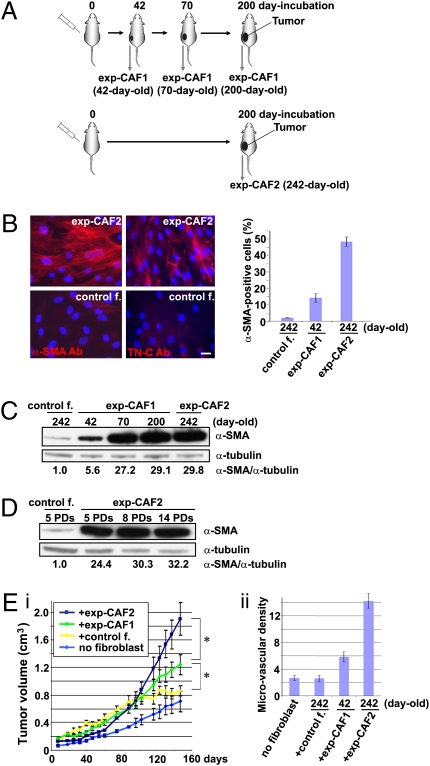
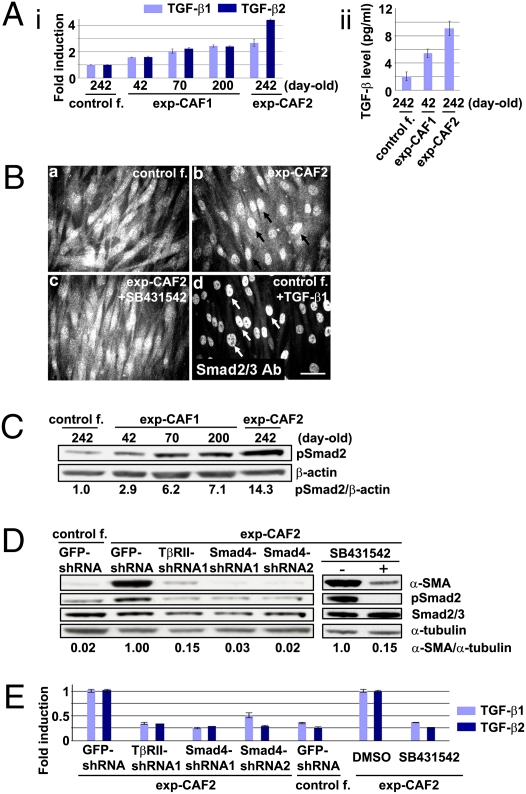
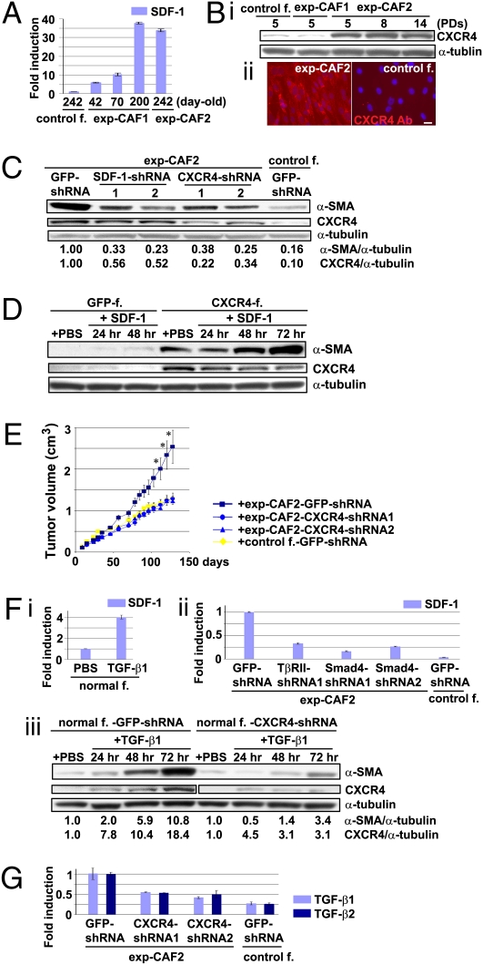
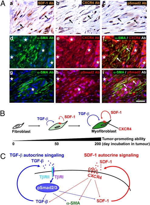
Much interest is currently focused on the emerging role of tumor-stroma interactions essential for supporting tumor progression. Carcinoma-associated fibroblasts (CAFs), frequently present in the stroma of human breast carcinomas, include a large number of myofibroblasts, a hallmark of activated fibroblasts. These fibroblasts have an ability to substantially promote tumorigenesis. However, the precise cellular origins of CAFs and the molecular mechanisms by which these cells evolve into tumor-promoting myofibroblasts remain unclear. Using a coimplantation breast tumor xenograft model, we show that resident human mammary fibroblasts progressively convert into CAF myofibroblasts during the course of tumor progression. These cells increasingly acquire two autocrine signaling loops, mediated by TGF-β and SDF-1 cytokines, which both act in autostimulatory and cross-communicating fashions. These autocrine-signaling loops initiate and maintain the differentiation of fibroblasts into myofibroblasts and the concurrent tumor-promoting phenotype. Collectively, these findings indicate that the establishment of the self-sustaining TGF-β and SDF-1 autocrine signaling gives rise to tumor-promoting CAF myofibroblasts during tumor progression. This autocrine-signaling mechanism may prove to be an attractive therapeutic target to block the evolution of tumor-promoting CAFs.


Conflict of interest statement

The authors declare no conflict of interest.

Figures

Fig. 1.
Fig. 1.
exp-CAFs mimic the tumor-promoting behavior of CAFs prepared from breast cancer patients. (A) Isolation of exp-CAFs. Normal GFP-labeled, puromycin-resistant, immortalized human mammary fibroblasts were coinjected with MCF-7-ras breast cancer cells s.c. into nude mice. Tumors were dissected at the indicated days and dissociated. The injected human fibroblasts were isolated under puromycin selection in culture and were termed exp-CAFs. See result for details. (B) Immunofluorescence of exp-CAF2 cells and control fibroblast-2 cells (control f.) to detect α-SMA (red) and tenascin-C (TN-C, red). Cell nuclei were stained with 4′,6-diamino-2-phenylindole (DAPI, blue). (Scale bar, 50 μm.) Error bars indicate SEM. (C and D) Western blotting of fibroblasts using an anti-α-SMA antibody. The membranes were also probed by an anti-α-tubulin antibody. The ratios of the signal intensity of α-SMA relative to α-tubulin are indicated. PDs, population doublings. (E) MCF-7-ras breast carcinoma cells were injected alone (n = 12) or coinjected with control fibroblast-2 cells (control f.; n = 10), 42-d-old exp-CAF1 cells (n = 12), or 242-d-old exp-CAF2 cells (n = 10) s.c. into nude mice. Tumor volume (i) was measured at the indicated days and microvascular density (ii) was quantified in tumors admixed with various fibroblasts 150 d after injection. *P < 0.05. Error bars, SE.
Fig. 2.
Fig. 2.
TGF-β autocrine signaling suffices to induce and maintain myofibroblast differentiation in exp-CAFs. (A, i) TGF-β1 and TGF-β2 mRNA expression in fibroblasts measured by real-time PCR. (ii) Active TGF-β concentrations in the media conditioned by fibroblasts measured by a luciferase assay. Error bars, SE. (B) Immunofluorescence for Smad2/3. Fibroblasts were incubated with control DMSO (a and b), SB431542, an inhibitor of the TGF-β type I receptor kinase (c), or recombinant TGF-β1 (d). Arrows (b, black; d, white) indicate nuclear Smad2/3 staining. (Scale bar, 100 μm.) (C) Western blotting of fibroblasts using anti-pSmad2 and anti-β-actin antibodies. The ratios of the signal intensity of pSmad2 relative to β-actin are indicated. (D) Western blotting of fibroblasts treated with the indicated shRNAs or SB431542 (10 μM). The membrane stripped was probed using different antibodies to detect α-SMA, pSmad2, Smad2/3, and α-tubulin. (E) Real-time PCR analysis of the fibroblasts described above. Error bars, SE.
Fig. 3.
Fig. 3.
SDF-1 autocrine signaling crosstalks with the TGF-β signaling to further boost myofibroblastic differentiation in exp-CAFs. (A) SDF-1 mRNA expression in fibroblasts measured by real-time PCR. Error bars, SE (B, i) Immunoblotting of fibroblasts using anti-CXCR4 and anti-α-tubulin antibodies. (ii) Immunofluorescence using an anti-CXCR4 antibody (red). Cell nuclei are stained with DAPI (blue). (Scale bar, 50 μm.) (C) Western blotting of fibroblasts expressing the indicated shRNAs to detect CXCR4. The membrane was also probed using antibodies against α-SMA and α-tubulin. The ratios of the signal intensity of either α-SMA or CXCR4 relative to α-tubulin are indicated. (D) Immunoblotting of human mammary fibroblasts expressing GFP or CXCR4 treated with or without SDF-1. (E) Exp-CAF2 (n = 12) or control fibroblast-2 (n = 10) cells expressing CXCR4- or GFP-shRNAs were injected with MCF-7-ras cells s.c. into nude mice. *P < 0.05. Error bars, SE. (F, i) Real-time PCR analysis of normal mammary fibroblasts treated with TGF-β1. (ii) Real-time PCR of fibroblasts treated with the indicated shRNAs. Error bars, SE. (iii) Western blotting of mammary fibroblasts expressing GFP- or CXCR4-shRNA treated with or without TGF-β1. The membrane stripped was probed with different antibodies to detect α-SMA, CXCR4, and α-tubulin. The observed CXCR4 signal in CXCR4-shRNA-expressing cells could only be detected using an enhanced chemiluminescence substrate. (G) Real-time PCR analysis of fibroblasts expressing CXCR4- or GFP-shRNA. Error bars, SE.
Fig. 4.
Fig. 4.
TGF-β and SDF-1 autocrine signaling operates in CAFs in invasive human breast carcinomas. (A) Immunohistochemistry of sections prepared from invasive human breast carcinomas using antibodies against SDF-1 (a, brown), CXCR4 (b, brown; e and f, red), pSmad2 (c, brown; h and i, red), and α-SMA (d, f, g, and i, green). The sections were also stained with hematoxylin (a–c, pale blue) or DAPI (d–i, blue). Cells staining positive are highlighted by arrows. A group of carcinoma cells is indicated by an asterisk. (Scale bar, 50 μm.) (B) During tumor progression, resident stromal fibroblasts within the tumor increasingly acquire two autocrine signaling loops involving TGF-β and SDF-1 that mediate transdifferentiation into tumor-promoting CAF myofibroblasts. (C) Schematic illustration describing two self-stimulating and cross-communicating signaling loops mediated by TGF-β and SDF-1 in CAF myofibroblasts. CAF-secreted TGF-β and SDF-1 ligands act upon TβR and CXCR4, respectively, in an autocrine fashion. The subsequent activation of TβR-Smad2/3 and CXCR4 signaling pathways drives myofibroblast differentiation and endogenous TGF-β and SDF-1 expression, thereby generating self-stimulating autocrine signaling loops that act in a feed-forward manner. Importantly, the TβR-Smad2/3 signaling also increases SDF-1 expression, thereby boosting SDF-1-CXCR4 autocrine signaling. This in turn elevates endogenous TGF-β expression. Cross-talk between these autocrine signaling loops therefore stimulates one another and further boosts myofibroblast differentiation in CAFs. A thick straight arrow indicates direct stimulatory modification, and thin straight arrows depict transcriptional contribution.

References

    1. Sappino AP, Skalli O, Jackson B, Schürch W, Gabbiani G. Smooth-muscle differentiation in stromal cells of malignant and non-malignant breast tissues. Int J Cancer. 1988;41:707–712. - PubMed
    1. Kellermann MG, et al. Mutual paracrine effects of oral squamous cell carcinoma cells and normal oral fibroblasts: Induction of fibroblast to myofibroblast transdifferentiation and modulation of tumor cell proliferation. Oral Oncol. 2008;44:509–517. - PubMed
    1. Cardone A, Tolino A, Zarcone R, Borruto Caracciolo G, Tartaglia E. Prognostic value of desmoplastic reaction and lymphocytic infiltration in the management of breast cancer. Panminerva Med. 1997;39:174–177. - PubMed
    1. Maeshima AM, et al. Modified scar grade: A prognostic indicator in small peripheral lung adenocarcinoma. Cancer. 2002;95:2546–2554. - PubMed
    1. Bissell MJ, Radisky D. Putting tumours in context. Nat Rev Cancer. 2001;1:46–54. - PMC - PubMed

Publication types

MeSH terms