Comparative Study
. 2010 Nov 23;107(47):20287-92.
doi: 10.1073/pnas.1011218107. Epub 2010 Nov 1.

Structure of the bacteriophage T4 long tail fiber receptor-binding tip

Affiliations
Comparative Study

Structure of the bacteriophage T4 long tail fiber receptor-binding tip

Sergio G Bartual et al. Proc Natl Acad Sci U S A. .

Abstract

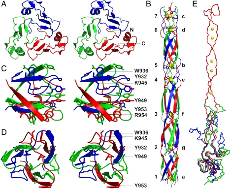
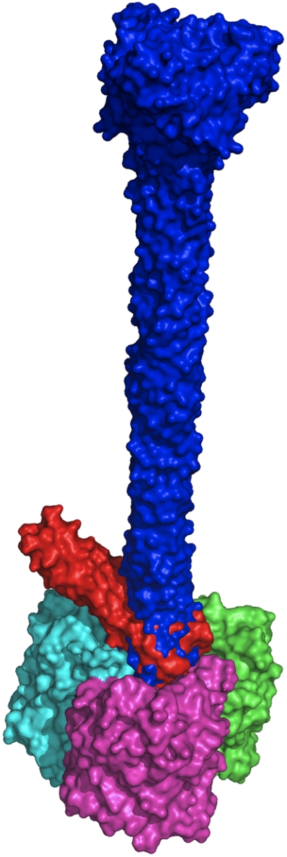
Bacteriophages are the most numerous organisms in the biosphere. In spite of their biological significance and the spectrum of potential applications, little high-resolution structural detail is available on their receptor-binding fibers. Here we present the crystal structure of the receptor-binding tip of the bacteriophage T4 long tail fiber, which is highly homologous to the tip of the bacteriophage lambda side tail fibers. This structure reveals an unusual elongated six-stranded antiparallel beta-strand needle domain containing seven iron ions coordinated by histidine residues arranged colinearly along the core of the biological unit. At the end of the tip, the three chains intertwine forming a broader head domain, which contains the putative receptor interaction site. The structure reveals a previously unknown beta-structured fibrous fold, provides insights into the remarkable stability of the fiber, and suggests a framework for mutations to expand or modulate receptor-binding specificity.


Conflict of interest statement

The authors declare no conflict of interest.

Figures

Fig. 1.
Fig. 1.
Bacteriophage T4 and its long tail fibers. (A and B) Schematic representations of bacteriophage T4 attached to a bacterial membrane before (Left) and after (Right) contraction of the outer tail tube. The tip domain of gp37 is boxed. (C) Schematic representation of the bacteriophage T4 long tail fiber. Domains P1–5 correspond to gp34; the kneecap domain (K-C) is formed by gp35, whereas the distal part of the fiber, consisting of gp36 and gp37, is divided into regions D1–11. The expressed protein, gp37(651–1026), corresponds to D9-11 (larger gray box), whereas the crystallized fragment, gp37(785–1026), corresponds to D10–11 (smaller gray box).
Fig. 2.
Fig. 2.
Structure of gp37(785–1026). Chains A, B, and C are colored red, green, and blue, respectively; iron ions are shown in yellow. (A) Ribbon representation. The N and C termini and every 10th residue of chain A are labeled. (B and C) Surface representations of the structure of gp37(785–1026) seen from the side (B) and top (C) to illustrate the extensive intertwining of the three protein chains in the trimer; domains are indicated; i.r. is the intertwined region between the collar and needle domains.
Fig. 3.
Fig. 3.
Domain structure of gp37(785–1026). (A) Walleyed stereo representation of the collar domain viewed from the direction of the needle domain. (B) The needle domain viewed from the side. Iron ions are represented as yellow balls. The histidine doublets coordinating the iron ions are shown and labeled with letters a-g; iron ions with numbers 1–7. (C and D) Side (C) and top (D) view of the head domain. Aromatic and basic side chains are shown and labeled (R954 is hidden behind Y953 in D). (E) Comparison of gp37(811–1026) with the bacteriophage T4 baseplate proteins gp12 and gp10. Superposition of monomers of gp37(811–1026) in red, onto gp12(330–527) in blue, and gp10 in green. Iron ions belonging to the gp37(811–1026) structure are shown in yellow, whereas the zinc ion identified in gp12 is shown in gray.
Fig. 4.
Fig. 4.
Alignment of the tip domains of the long tail fiber gp37 proteins of bacteriophages T4 (UniProt code P03744), TuIa (S13237), and TuIb (S13239) and of the side tail fiber of bacteriophage lambda (P03764). Of the latter two, only the sequence of the C-terminal 382 and 267 amino acids are known, respectively, although their entire gp37 proteins are expected to be similar in size to gp37 of T4. Identical residues are indicated with asterisks and similar ones with dots. Hydrophobic residues contributing to the central longitudinal core of the needle domain are boxed in gray; His-X-His motifs are also labeled with letters on top of the alignment. A deletion of 10 amino acids in the T4 needle domain after residue 909 with respect to the others is compensated for by a deletion of nine amino acids just before residue 979 in the “return” strand; these last nine amino acids contain a putative eighth metal-binding site His-Ala-His for TuIa, TuIb, and lambda.
Fig. 5.
Fig. 5.
Docking of gp37 into outer membrane porin protein C. Two superimposed results of automatic docking experiments are shown with the gp37(811–1026) trimer in blue, a truncated trimeric gp37(918–973) model in red, and the three subunits of the outer membrane porin protein C trimer (PDB code 2J1N) in green, magenta, and cyan.

References

    1. Hagens S, Loessner MJ. Application of bacteriophages for detection and control of foodborne pathogens. Appl Microbiol Biotechnol. 2007;76:513–519. - PubMed
    1. Petrenko VA, Vodyanoy VJ. Phage display for detection of biological threat agents. J Microbiol Methods. 2003;53:253–262. - PubMed
    1. Parisien A, Allain B, Zhang J, Mandeville R, Lan CQ. Novel alternatives to antibiotics: Bacteriophages, bacterial cell wall hydrolases, and antimicrobial peptides. J Appl Microbiol. 2008;104:1–13. - PubMed
    1. Chanishvili N, Sharp RA. Literature Review of the Practical Application of Bacteriophage Research. Tbilisi, Georgia: Eliava Inst of Bacteriophage, Microbiology and Virology; 2009.
    1. Wright A, Hawkins CH, Anggard EE, Harper DR. A controlled clinical trial of a therapeutic bacteriophage preparation in chronic otitis due to antibiotic-resistant Pseudomonas aeruginosa; a preliminary report of efficacy. Clin Otolaryngol. 2009;34:349–357. - PubMed

Publication types

Substances

Associated data

LinkOut - more resources