. 2010 Nov 16;122(20):2039-47.
doi: 10.1161/CIRCULATIONAHA.109.930222. Epub 2010 Nov 1.

Titin is a target of matrix metalloproteinase-2: implications in myocardial ischemia/reperfusion injury

Affiliations

Titin is a target of matrix metalloproteinase-2: implications in myocardial ischemia/reperfusion injury

Mohammad A M Ali et al. Circulation. .

Abstract

Background: Titin is the largest mammalian (≈3000 to 4000 kDa) and myofilament protein that acts as a molecular spring in the cardiac sarcomere and determines systolic and diastolic function. Loss of titin in ischemic hearts has been reported, but the mechanism of titin degradation is not well understood. Matrix metalloproteinase-2 (MMP-2) is localized to the cardiac sarcomere and, on activation in ischemia/reperfusion injury, proteolyzes specific myofilament proteins. Here we determine whether titin is an intracellular substrate for MMP-2 and if its degradation during ischemia/reperfusion contributes to cardiac contractile dysfunction.

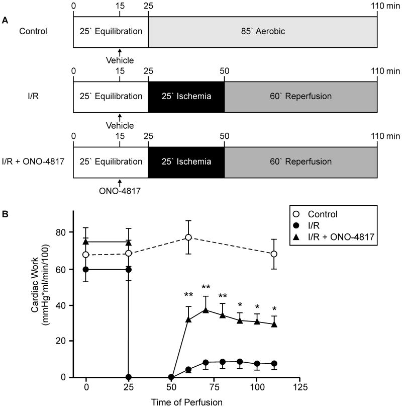
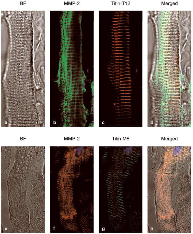
Methods and results: Immunohistochemistry and confocal microscopy in rat and human hearts showed discrete colocalization between MMP-2 and titin in the Z-disk region of titin and that MMP-2 is localized mainly to titin near the Z disk of the cardiac sarcomere. Both purified titin and titin in skinned cardiomyocytes were proteolyzed when incubated with MMP-2 in a concentration-dependent manner, and this was prevented by MMP inhibitors. Isolated rat hearts subjected to ischemia/reperfusion injury showed cleavage of titin in ventricular extracts by gel electrophoresis, which was confirmed by reduced titin immunostaining in tissue sections. Inhibition of MMP activity with ONO-4817 prevented ischemia/reperfusion-induced titin degradation and improved the recovery of myocardial contractile function. Titin degradation was also reduced in hearts from MMP-2 knockout mice subjected to ischemia/reperfusion in vivo compared with wild-type controls.

Conclusion: MMP-2 localizes to titin at the Z-disk region of the cardiac sarcomere and contributes to titin degradation in myocardial ischemia/reperfusion injury.


Figures

Figure 1
Figure 1
Co-localization of MMP-2 and titin at the Z disc region of the left ventricular cardiac sarcomere in 10 min aerobically perfused rat hearts (longitudinal sections). MMP-2 shows better co-localization with T12 than M8 epitope of titin in the sarcomere of left ventricular myocardium. MMP-2-immunoreactivity reveals at Z-lines with high density as well as M-lines with low density. T12 epitope reveals at only Z-lines and M8 epitope reveals at only M-lines. a-c shows that high density of MMP-2 (green) co-localizes (yellow) with T12 epitope (red) at the Z-lines. d-f shows that low density of MMP-2 (red) co-localizes (yellow) with M8 epitope (green) at M-lines. Scale bar is 5 μm for all images except for the enlarged portion of c illustrating the Z- and M-lines.
Figure 2
Figure 2
In vitro incubation (60 min, 37°C) of skinned cardiomyocytes with MMP-2. A, MMP-2 cleaved cardiac titin (T1) in a concentration-dependent manner (4-120 nmol/L) as shown by the appearance of the titin degradation product (T2). B, The cleavage of titin by MMP-2 was prevented by inhibiting the activity of MMP-2 with MMPs inhibitors o-phenanthroline (o-ph) or ONO-4817. T2/T1 band density ratio is indicated below each lane. MHC (myosin heavy chain) as loading control.
Figure 3
Figure 3
Mechanical recovery of isolated perfused working rat hearts subjected to 25 min global, no-flow ischemia followed by 60 min reperfusion without (I/R) or with 50 μmol/L ONO-4817 (I/R + ONO-4817) in comparison with aerobically perfused control hearts. A, Schematic representation of the perfusion protocols for Control (n=6), I/R (n=7) and I/R + ONO-4817 (n=8) groups. B, Time course of changes in cardiac work of isolated working rat hearts. **p<0.001, *p<0.05 versus corresponding values of I/R group, repeated measures two-way ANOVA.
Figure 4
Figure 4
Titin degradation in ischemic-reperfused rat hearts. A, Representative SDS-agarose gel for analysis of titin in ventricular extracts. Titin (T1) and titin degradation product (T2) in ventricular homogenates from control, I/R and I/R + ONO-4817 hearts analyzed using a 1% vertical SDS-agarose gel. MHC (myosin heavy chain). BLV (bovine left ventricle) was used as a standard and shows N2BA and N2B isoforms of titin; note that the majority of rat heart titin is the N2B isoform. Each lane is an extract from individually perfused hearts. B, Ratio of total titin (T1 + T2) to MHC content (n=6 in each group). C, Ratio of T2 titin to MHC content (n=6 in each group). *p<0.05 versus control (One-way ANOVA, Tukey's post hoc test). D, Representative left ventricular cryosections immunostained against titin epitope 9D10. Titin immunostaining using the 9D10 antibody (raised against the proline-glutamate-valine-lysine, PEVK, domain) was decreased in I/R hearts compared to control, whereas staining intensity was comparable between I/R + ONO-4817 and control hearts. Scale bar is 10 μm for all images. Images are representative of at least four individual hearts investigated under each condition.
Figure 5
Figure 5
Titin degradation in MMP-2 knockout (KO) and wild type (WT) mouse hearts subjected to ischemia/reperfusion (I/R) in vivo. A, Representative 1% vertical SDS-agarose gel shows titin (T1) isoforms (N2BA and N2B) and titin degradation product T2 in left ventricle from Sham or in ischemic regions from I/R groups in either wild type or MMP-2 knockout mice. B, Quantification of T2 titin to total titin ratios (n=6 in each group). *p<0.01 versus Sham control (One-way ANOVA, Tukey's post hoc test).
Figure 6
Figure 6
Co-localization of MMP-2 and titin near the Z-disc in diseased human heart. Left ventricle sections were used from the explanted failing heart from a patient receiving a heart transplant. a-d shows that high density of MMP-2 (green) co-localizes (yellow) with T12 epitope (red) at the Z-lines. e-h shows that low density of MMP-2 (red) co-localizes (yellow) with M8 epitope (green) at M-lines. BF (bright field images). Scale bar is 10 μm.

Comment in

References

    1. Birkedal-Hansen H. Proteolytic remodeling of extracellular matrix. Curr Opin Cell Biol. 1995;7:728–735. - PubMed
    1. Yasmin W, Strynadka KD, Schulz R. Generation of peroxynitrite contributes to ischemia-reperfusion injury in isolated rat hearts. Cardiovasc Res. 1997;33:422–432. - PubMed
    1. Okamoto T, Akaike T, Sawa T, Miyamoto Y, van der Vliet A, Maeda H. Activation of matrix metalloproteinases by peroxynitrite-induced protein S-glutathiolation via disulfide S-oxide formation. J Biol Chem. 2001;276:29596–29602. - PubMed
    1. Viappiani S, Nicolescu AC, Holt A, Sawicki G, Crawford BD, Leon H, van Mulligen T, Schulz R. Activation and modulation of 72kDa matrix metalloproteinase-2 by peroxynitrite and glutathione. Biochem Pharmacol. 2009;77:826–834. - PubMed
    1. Woessner JF., Jr Matrix metalloproteinases and their inhibitors in connective tissue remodeling. FASEB J. 1991;5:2145–2154. - PubMed

Publication types

MeSH terms