. 2010 Nov 16;122(20):2031-8.
doi: 10.1161/CIRCULATIONAHA.109.866053. Epub 2010 Nov 1.

Magnetic resonance imaging of carotid atherosclerotic plaque in clinically suspected acute transient ischemic attack and acute ischemic stroke

Affiliations

Magnetic resonance imaging of carotid atherosclerotic plaque in clinically suspected acute transient ischemic attack and acute ischemic stroke

Jaywant P Parmar et al. Circulation. .

Abstract

Background: Carotid atherosclerotic plaque rupture is thought to cause transient ischemic attack (TIA) and ischemic stroke (IS). Pathological hallmarks of these plaques have been identified through observational studies. Although generally accepted, the relationship between cerebral thromboembolism and in situ atherosclerotic plaque morphology has never been directly observed noninvasively in the acute setting.

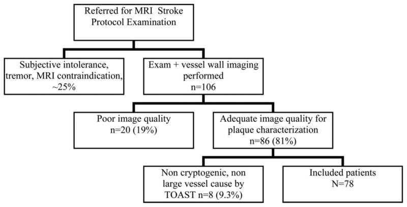
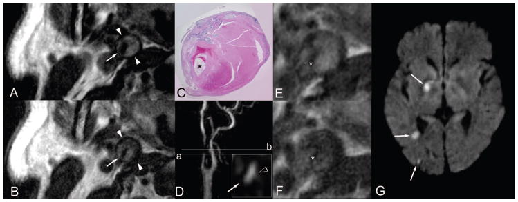
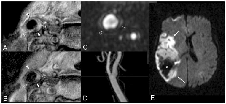
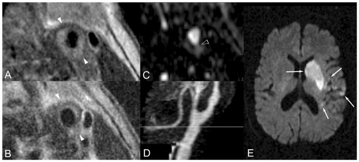
Methods and results: Consecutive acutely symptomatic patients referred for stroke protocol magnetic resonance imaging/angiography underwent additional T1- and T2-weighted carotid bifurcation imaging with the use of a 3-dimensional technique with blood signal suppression. Two blinded reviewers performed plaque gradings according to the American Heart Association classification system. Discharge outcomes and brain magnetic resonance imaging results were obtained. Image quality for plaque characterization was adequate in 86 of 106 patients (81%). Eight TIA/IS patients with noncarotid pathogenesis were excluded, yielding 78 study patients (38 men and 40 women with a mean age of 64.3 years, SD 14.7) with 156 paired watershed vessel/cerebral hemisphere observations. Thirty-seven patients had 40 TIA/IS events. There was a significant association between type VI plaque (demonstrating cap rupture, hemorrhage, and/or thrombosis) and ipsilateral TIA/IS (P<0.001). A multiple logistic regression model including standard Framingham risk factors and type VI plaque was constructed. Type VI plaque was the dominant outcome-associated observation achieving significance (P<0.0001; odds ratio, 11.66; 95% confidence interval, 5.31 to 25.60).

Conclusions: In situ type VI carotid bifurcation region plaque identified by magnetic resonance imaging is associated with ipsilateral acute TIA/IS as an independent identifier of events, thereby supporting the dominant disease pathophysiology.


Figures

Figure 1
Figure 1
Eligibility of the subjects included in the analysis. TOAST indicates Trial of Org 10172 in Acute Stroke Treatment.
Figure 2
Figure 2
A through G, Stenotic, ruptured, hemorrhagic/thrombotic type VI atherosclerotic plaque and acute embolic stroke. A 63-year-old man presented with acute onset of left-sided weakness, left neglect, and associated “blackouts.” He was imaged within 48 hours of symptom onset and underwent emergency carotid endarterectomy at 70 hours. A and B, T1- and T2-weighted blood-suppressed MR images demonstrate a large burden of internal carotid artery plaque. Axial sections A and B were obtained at a level indicated by the reference line labeled (a) on D, a maximum intensity projection of the MRA. The inset in image D illustrates axial MRA data at the level (a), with the hollow white arrowhead indicating a focal fading of the contrast column into the vessel wall compatible with the leading edge of a cap rupture. The lumen is indicated by a fine white arrow on A, B, and D (inset). C, E, and F demonstrate a magnification ×10 hematoxylineosin–stained histological section of the endarterectomy specimen in the region of occlusion and corresponding axial T1- and T2-weighted images, all correlating with the reference line labeled (b) on MRA projection (D). These images demonstrate type VI atherosclerotic plaque comprised of hemorrhagic components in various stages of organization, with the lumen indicated (*). Fresh blood is demonstrated in the paraluminal 12 o’clock position, with organizing red and white thrombus demonstrated ranging from the paraluminal 3 (most organized) through 6 o’clock (least organized) positions. Chronic and fibrotic organized thrombus occupies the peripheral wall spanning the 1 through 4 o’clock positions. Calcification is distributed in the extreme peripheral wall circumferentially ranging from the 11 through 3 o’clock positions. The patient suffered multiple acute strokes in the right carotid watershed as demonstrated on the clinical cerebral diffusion-weighted image (G).
Figure 3
Figure 3
A through E, Nonstenotic hemorrhagic, thrombotic type VI atherosclerotic plaque, and acute middle cerebral artery territory hemorrhagic stroke. A 77-year-old woman with hypertension, untreated hyperlipidemia, and a current cigarette smoking habit presented with left-sided weakness and mental status changes and was imaged 8 hours from onset. A and B, T1- and T2-weighted axial blood-suppressed MR images demonstrate an abnormal eccentrically thickened right common carotid artery wall as indicated ranging between hollow arrowheads. Radial signal inhomogeneity in this region suggests type VI atherosclerotic plaque. C is axially reformatted 3D MRA data. D is a maximum intensity projection of those data simulating a projectional arteriogram demonstrating no significant luminal stenosis. A reference line indicating the level of A, B, and C is demonstrated. The region of greatest plaque volume as identified on A and B and demarcated between hollow arrowheads on A, B, and C shows heterogeneous intrinsic signal intensity compatible with intramural hemorrhage with a geographically associated focal region of endoluminal irregular contour on C indicating overlying thrombosis in situ and securing the overall classification of type VI. Image E is the clinical cerebral diffusion-weighted image that demonstrates a large acute right hemisphere middle cerebral artery territory hemorrhagic infarction (*).
Figure 4
Figure 4
A through E, Nonstenotic ruptured, hemorrhagic type VI plaque and acute embolic stroke. A 79-year-old man with hypertension presented with acute onset right-sided numbness, weakness, and inability to speak. He was imaged within 28 hours of symptom onset. A and B, T1- and T2-weighted blood-suppressed images demonstrate an abnormally thickened vessel wall with radial signal inhomogeneity suggesting type VI atherosclerotic plaque. C, Axially reformatted 3D MRA data demonstrate contrasted blood extending into the vessel wall as indicated by the arrowhead, tracking circumferentially between the 4 and 8 o’clock positions, a specific sign indicating plaque cap rupture and thereby confirming the type VI classification. The MRA maximum intensity projection (D) demonstrates a focal mild circumferential stenosis at the level of this plaque, indicated by the gray line. A representative section of the clinical cerebral diffusion-weighted image (E) demonstrates multiple acute left middle cerebral artery territory infarctions, indicated by white arrows.

References

    1. Executive Committee for the Asymptomatic Carotid Atherosclerosis Study. Endarterectomy for asymptomatic carotid artery stenosis. JAMA. 1995;273:1421–1428. - PubMed
    1. North American Symptomatic Carotid Endarterectomy Trial Collaborators. Beneficial effect of carotid endarterectomy in symptomatic patients with high-grade carotid stenosis. N Engl J Med. 1991;325:445–453. - PubMed
    1. European Carotid Surgery Trialists’ Collaborative Group. MRC European Carotid Surgery Trial: interim results for symptomatic patients with severe (70–99%) or with mild (0–29%) carotid stenosis. Lancet. 1991;337:1235–1243. - PubMed
    1. Glagov S, Weisenberg E, Zarins CK, Stankunavicius R, Kolettis GJ. Compensatory enlargement of human atherosclerotic coronary arteries. N Engl J Med. 1987;316:1371–1375. - PubMed
    1. Ambrose JA, Tannenbaum MA, Alexopoulos D, Hjemdahl-Monsen CE, Leavy J, Weiss M, Borrico S, Gorlin R, Fuster V. Angiographic progression of coronary artery disease and the development of myocardial infarction. J Am Coll Cardiol. 1988;12:56–62. - PubMed

Publication types

MeSH terms