Comparative Study
. 2010 Nov;66(Pt 11):1153-63.
doi: 10.1107/S0907444910026582. Epub 2010 Oct 20.

Joint X-ray and neutron refinement with phenix.refine

Affiliations
Comparative Study

Joint X-ray and neutron refinement with phenix.refine

Pavel V Afonine et al. Acta Crystallogr D Biol Crystallogr. 2010 Nov.

Abstract

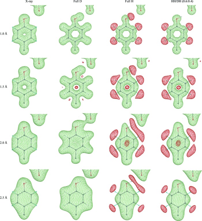
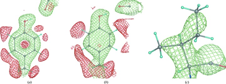
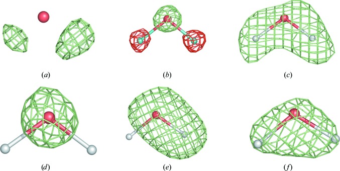
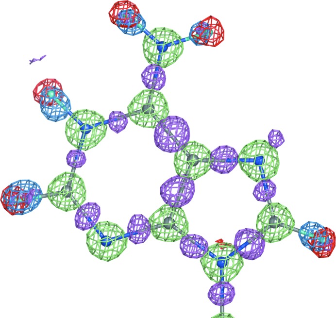
Approximately 85% of the structures deposited in the Protein Data Bank have been solved using X-ray crystallography, making it the leading method for three-dimensional structure determination of macromolecules. One of the limitations of the method is that the typical data quality (resolution) does not allow the direct determination of H-atom positions. Most hydrogen positions can be inferred from the positions of other atoms and therefore can be readily included into the structure model as a priori knowledge. However, this may not be the case in biologically active sites of macromolecules, where the presence and position of hydrogen is crucial to the enzymatic mechanism. This makes the application of neutron crystallography in biology particularly important, as H atoms can be clearly located in experimental neutron scattering density maps. Without exception, when a neutron structure is determined the corresponding X-ray structure is also known, making it possible to derive the complete structure using both data sets. Here, the implementation of crystallographic structure-refinement procedures that include both X-ray and neutron data (separate or jointly) in the PHENIX system is described.


Figures

Figure 1
Figure 1
(F calc, ϕcalc) Fourier syntheses computed at different resolutions (1, 1.5, 2 and 2.5 Å) and corresponding to four different cases: X-ray, neutron fully deuterated, neutron fully hydrogenated and neutron partially deuterated, in which the hydroxyl H (HH) of tyrosine shares its site with deuterium (DH) with an occupancy ratio of 0.6:0.4.
Figure 2
Figure 2
Some typical 2F obsDF model nuclear map appearances. (a) Tyr12 of 1iu5 (+1.2σ and −1.5σ, 1.6 Å resolution), (b) Tyr92 (+1.2σ and −1.4σ) and (c) Ile81 (+2.0σ) of 5rsa (2.0 Å resolution).
Figure 3
Figure 3
A water molecule in (a) an mF obsDF model X-ray map at 0.66 Å resolution and (b) a 2F obsDF model nuclear map at 0.65 Å resolution; the remaining four nuclear maps are computed at 1.5 Å resolution.
Figure 4
Figure 4
Three maps overlaid with a fragment of the NAD structure (UR0013): 2mF obsDF model neutron map (shown at ±2.4σ; positive and negative contours are shown in lime and red, respectively; computed at 0.65 Å resolution) and two mF obsDF model maps (X-ray; computed at 0.6 Å resolution; blue is contoured at 5σ and computed with H atoms omitted, magenta is contoured at 2.8σ and computed with all atoms included). Both structures, X-ray and neutron, were re-refined in phenix.refine to R factors (R work) of 2.09% and 7.97%, respectively. Red (negative) density corresponds to the hydrogen nucleus positions, blue density shows the shifted concentrations of hydrogen’s electron density revealed by X-rays, magenta shows the bonding electron density and lime is positive nuclear density.
Figure 5
Figure 5
Averaged ΔR work and ΔR free as a function of resolution computed for structures refined with and without H atoms added. See text for details.
Figure 6
Figure 6
2mF obsDF model nuclear map for the NAD structure (UR0013) contoured at ±2.4σ; lime is positive and red is negative.
Figure 7
Figure 7
2mF obsDF model nuclear map: orientation of an O—H/D bond before (wrong, left) and after (correct, right) automatic real-space correction performed during refinement in phenix.refine.

References

    1. Adams, P. D. et al. (2010). Acta Cryst. D66, 213–221.
    1. Adams, P. D., Mustyakimov, M., Afonine, P. V. & Langan, P. (2009). Acta Cryst. D65, 567–573. - PMC - PubMed
    1. Adams, P. D., Pannu, N. S., Read, R. J. & Brünger, A. T. (1997). Proc. Natl Acad. Sci. USA, 94, 5018–5023. - PMC - PubMed
    1. Afonine, P. V., Grosse-Kunstleve, R. W. & Adams, P. D. (2005a). Acta Cryst. D61, 850–855. - PMC - PubMed
    1. Afonine, P. V., Grosse-Kunstleve, R. W. & Adams, P. D. (2005b). CCP4 Newsl. 42, contribution 8.

Publication types

MeSH terms