Review
. 2010 Nov;120(11):3779-87.
doi: 10.1172/JCI43766. Epub 2010 Nov 1.

Central modulation of pain

Affiliations
Review

Central modulation of pain

Michael H Ossipov et al. J Clin Invest. 2010 Nov.

Abstract

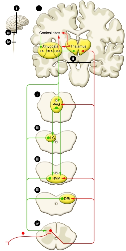
It has long been appreciated that the experience of pain is highly variable between individuals. Pain results from activation of sensory receptors specialized to detect actual or impending tissue damage (i.e., nociceptors). However, a direct correlation between activation of nociceptors and the sensory experience of pain is not always apparent. Even in cases in which the severity of injury appears similar, individual pain experiences may vary dramatically. Emotional state, degree of anxiety, attention and distraction, past experiences, memories, and many other factors can either enhance or diminish the pain experience. Here, we review evidence for "top-down" modulatory circuits that profoundly change the sensory experience of pain.


Figures

Figure 1
Figure 1. Schematic representation of pain modularity circuitry.
Nociceptive inputs enter the spinal dorsal horn through primary afferent fibers that synapse onto transmission neurons. The projection fibers ascend through the contralateral spinothalamic tract. Ascending projections target the thalamus, and collateral projections also target mesencephalic nuclei, including the DRt, the RVM, and the midbrain PAG. Descending projections from the DRt are a critical component of the DNIC pathway. Rostral projections from the thalamus target areas that include cortical sites and the amygdala. The lateral capsular part of the CeA (“nociceptive amygdala”) receives nociceptive inputs from the brainstem and spinal cord. Inputs from the thalamus and cortex enter through the lateral (LA) and basolateral (BLA) amygdala. The CeA sends outputs to cortical sites and the thalamus, in which cognitive and conscious perceptions of pain are integrated. Descending pain modulation is mediated through projections to the PAG, which also receives inputs from other sites, including the hypothalamus (data not shown), and communicates with the RVM as well as other medullary nuclei that send descending projections to the spinal dorsal horn through the DLF. The noradrenergic locus coeruleus (LC) receives inputs from the PAG, communicates with the RVM, and sends descending noradrenergic inhibitory projections to the spinal cord. Antinociceptive and pronociceptive spinopetal projections from the RVM positively and negatively modulate nociceptive inputs and provide for an endogenous pain regulatory system. Ascending (red) and descending (green) tracts are shown schematically. Areas labeled “i–iv” in the small diagram correspond with labeled details of the larger diagram.
Figure 2
Figure 2. Schematic representation of bulbospinal pain inhibition and potential targets of analgesic activity.
(A) Descending pain inhibition from the PAG can be initiated by electrical stimulation or direct microinjection of opioids. Recent evidence also indicates a role for COX inhibitors in the PAG as well. Opioids and cannabinoids inhibit pain by enhancing the baseline firing rate of off-cells and eliminating the off-cell pause in response to nociceptive stimuli. Inhibition of on-cell activity may abolish enhanced pain states. The on-cells and off-cells might correlate with pain facilitatory (+) and inhibitory (–) neurons in the RVM, respectively. At the level of the spinal cord, opioids can inhibit transmitter release from primary afferent terminals as well as activity of pain transmission neurons. Norepinephrine (NE) release from spinopetal noradrenergic fibers from medullary sites also inhibits pain transmission. Tricyclic antidepressants (TCAs) and other norepinephrine reuptake inhibitors enhance the antinociceptive effect of opioids by increasing the availability of spinal norepinephrine (box). Areas labeled “i–iii” in the small diagram correspond with labeled details of the larger diagram. α2A, α2-adrenergic receptor; DRG, dorsal root ganglion; SNRI, serotonin/norepinephrine reuptake inhibitor; SP, substance P. (B) Mice deficient in dopamine β-hydroxylase that do not produce norepinephrine show a diminished antinociceptive effect of morphine compared with control animals, suggesting that the presence of norepinephrine, presumably released in the spinal cord, is required for the full expression of morphine antinociception. The dashed line represents the 50% effect, and the corresponding dose is the ED50 (that is the dose producing a 50% effect). % MPE, percentage maximal possible effect. ***P < 0.001 compared with the control group. Error bars represent SEM. Copyright National Academy of Sciences, USA (151).

References

    1. Beecher HK. Pain in men wounded in battle. Ann Surg. 1946;123(1):96–105. doi: 10.1097/00000658-194601000-00008. - DOI - PMC - PubMed
    1. Bingel U, Tracey I. Imaging CNS modulation of pain in humans. Physiology (Bethesda). 2008;23(3):371–380. - PubMed
    1. Fields HL, Basbaum AI, Heinricher MM. Central nervous system mechanisms of pain modulation. In: McMahon S, Koltzenburg M, eds.Textbook of Pain . 5th ed. Burlington, Massachusetts, USA: Elsevier Health Sciences; 2005:125–142.
    1. Beecher HK. The powerful placebo. J Am Med Assoc. 1955;159(17):1602–1606. - PubMed
    1. Levine JD, Gordon NC, Fields HL. The mechanism of placebo analgesia. Lancet. 1978;2(8091):654–657. - PubMed

Publication types