. 2010 Oct 21;5(10):e13557.
doi: 10.1371/journal.pone.0013557.

Multiple FadD acyl-CoA synthetases contribute to differential fatty acid degradation and virulence in Pseudomonas aeruginosa

Affiliations

Multiple FadD acyl-CoA synthetases contribute to differential fatty acid degradation and virulence in Pseudomonas aeruginosa

Yun Kang et al. PLoS One. .

Abstract

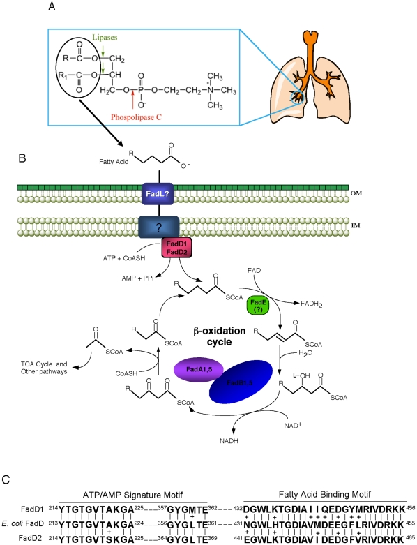
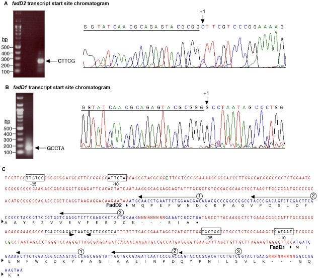
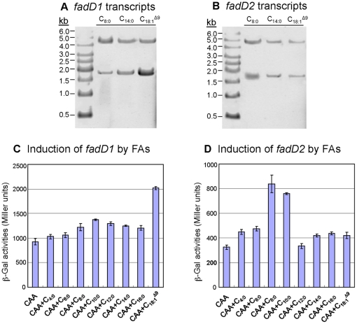
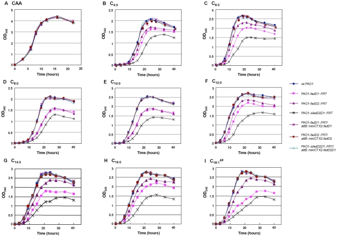
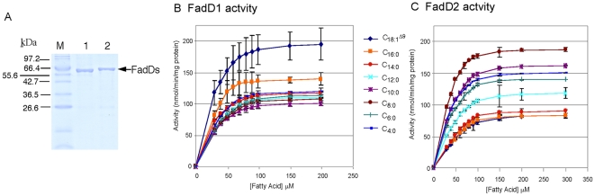
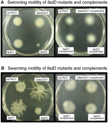
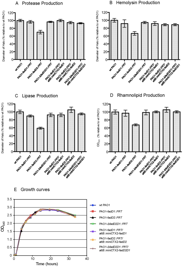
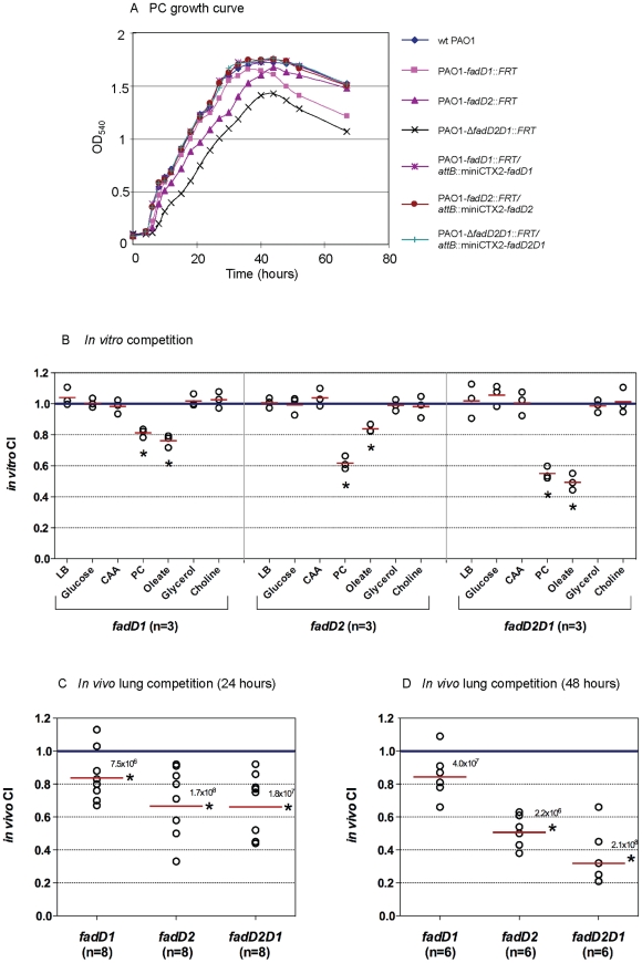
A close interconnection between nutrient metabolism and virulence factor expression contributes to the pathophysiology of Pseudomonas aeruginosa as a successful pathogen. P. aeruginosa fatty acid (FA) degradation is complicated with multiple acyl-CoA synthetase homologs (FadDs) expressed in vivo in lung tissue during cystic fibrosis infections. The promoters of two genetically linked P. aeruginosa fadD genes (fadD1 and fadD2) were mapped and northern blot analysis indicated they could exist on two different transcripts. These FadDs contain ATP/AMP signature and FA-binding motifs highly homologous to those of the Escherichia coli FadD. Upon introduction into an E. coli fadD(-)/fadR(-) double mutant, both P. aeruginosa fadDs functionally complemented the E. coli fadD(-)/fadR(-) mutant, allowing degradation of different chain-length FAs. Chromosomal mutagenesis, growth analysis, induction studies, and determination of kinetic parameters suggested that FadD1 has a substrate preference for long-chain FAs while FadD2 prefers shorter-chain FAs. When compared to the wild type strain, the fadD2 mutant exhibited decreased production of lipase, protease, rhamnolipid and phospholipase, and retardation of both swimming and swarming motilities. Interestingly, fadD1 mutant showed only increased swarming motility. Growth analysis of the fadD mutants showed noticeable deficiencies in utilizing FAs and phosphatidylcholine (major components of lung surfactant) as the sole carbon source. This defect translated into decreased in vivo fitness of P. aeruginosa in a BALB/c mouse lung infection model, supporting the role of lipids as a significant nutrient source for this bacterium in vivo.


Conflict of interest statement

Competing Interests: The authors have declared that no competing interests exist.

Figures

Figure 1
Figure 1. The proposed FA degradation pathway in P. aeruginosa based on E. coli β-oxidation.
(A) Phosphatidylcholine (PC) is the major component of lung surfactant. PC can be cleaved by P. aeruginosa phospholipase C and lipases producing free fatty acids that are degraded via the β-oxidation pathway by this bacterium. (B) FAs are transported through the outer membrane aided by an unidentified P. aeruginosa FadL , . In E. coli, FA may be transported through the inner membrane via an unknown mechanism coupled to a single peripheral membrane FadD protein . However, P. aeruginosa contains at least two FadDs (FadD1 and FadD2). Although there are over a dozen potential FadE homologues in the P. aeruginosa genome, the specific enzyme(s) that catalyzes this reaction has not been identified. FadB catalyzes the next two steps followed by cleavage of the 3-keto-acyl-CoA by FadA. Two fadBA operons (fadBA1 and fadBA5) have been identified in P. aeruginosa , . (C) Alignment of the P. aeruginosa FadD1 and FadD2 ATP/AMP-signature and FA-binding motifs with the FadD motifs of E. coli , . Abbreviation for Fad-proteins: FadA, 3-ketoacyl-CoA thiolase; FadB, enoyl-CoA hydratase and 3-hydroxyacyl-CoA dehydrogenase; FadD, acyl-CoA synthetase; FadE, acyl-CoA dehydrogenase; FadL, outer membrane FA translocase.
Figure 2
Figure 2. SMART mapping of the transcriptional start sites for fadD2 and fadD1.
(A) One SMART product was observed after PCR amplification of the cDNA with SMART and fadD2 primers (oligonucleotides #798 and #373). Sequencing of the single band with a nested fadD2 primer (oligonucleotide #374) displayed a reverse-complement sequence chromatogram, showing the fadD2 transcriptional start site (indicated by +1 at the CTTCG sequence) and the underlined SMART primer sequence. (B) Likewise, the downstream fadD1 transcriptional start site was mapped (at the G of the sequence GCCTA) by sequencing a single PCR product. (C) fadD2 and fadD1 coding sequences and the predicted −10 and −35 promoter regions are indicated (boxed). The intergenic region between fadD2 and fadD1 contains a potential transcriptional terminator or attenuator sequence (inverted arrows). For each gene, three black arrows indicate primers 1, 2 and 3 (#372/#375, #373/376, and #374/377) used for mapping fadD2 and fadD1. Dashed lines indicate missing protein sequences and dots indicate stop codons.
Figure 3
Figure 3. Transcriptional profile of fadD1 and fadD2 in various FAs.
For a short- (C8:0), medium- (C10:0), and long-chain FA (C18:1 Δ9), northern blot analysis indicated two possible transcripts for both fadD genes when probed with either fadD1 (A) or fadD2 (B). Gene-fusion studies of strains P518 (PfadD1-lacZ) and P520 (PfadD2-lacZ), grown to mid-log phase, showed differential expression of fadD1 and fadD2 in the presence of different FAs (C and D). (C) fadD1 was up-regulated in the presence of the unsaturated LCFA (C18:1 Δ9), while fadD2 expression was significantly increased in the presence of shorter chain FAs (C8:0, C10:0) (D). For (C) and (D), all cultures had identical growth-rates and overall cell densities (data not shown).
Figure 4
Figure 4. Growth analysis of fadD mutants using various FAs as sole carbon sources.
Although fadD mutants showed various defects when grown with FAs of different chain-lengths (see top of graphs in B-I), no growth defects were observed for any of the mutants when grown with casamino acids (CAA) as a control (A). All three P. aeruginosa mutants were fully complemented by the respective missing gene(s) and grew as well as the wildtype PAO1 on all carbon sources. Not shown are the three control mutant strains (PAO1-fadD1::FRT/attB::miniCTX2, PAO1-fadD2::FRT/attB::miniCTX2, and PAO1-ΔfadD2D1::FRT/attB::miniCTX2) having the empty miniCTX2 integrated into their chromosomes, where all had similar growth characteristics to the non-complemented mutants.
Figure 5
Figure 5. Purification and biochemical characterization of the two P. aeruginosa FadDs.
(A) FadD1 (lane 1; MW = 61,655) and FadD2 (lane 2; MW = 61,737) were purified to near homogeneity from an E. coli fadD - strain to prevent potential contamination of E. coli FadD. FadD1 activities (B) were higher for LCFAs (C18:1 Δ9>C16:0), while FadD2 (C) had higher activities for shorter chain FAs (C8:0>C10:0>C4:0>C6:0).
Figure 6
Figure 6. Altered swimming and swarming motility of P. aeruginosa fadD mutants.
(A) Swimming motility of fadD mutants and their complements. (B) Swarming migration of fadD mutants and their complements. These figures are representative of multiple experiments. Strain designation is the same as shown in Table 3: wildtype PAO1, P007; fadD1 -, P175; fadD2 -, P547; ΔfadD2D1, P177; fadD1 - complement, P541; fadD2 - complement, P549; and ΔfadD2D1 complement, P543.
Figure 7
Figure 7. Analysis of protease, hemolysin, lipase, and rhamnolipid production by P. aeruginosa fadD mutants.
The fadD2 mutant displayed significantly decreased production of proteases (A), hemolysins (B), lipases (C), and rhamnolipids (D), while no growth defects in LB were observed (E). These assays were conducted in triplicate and are expressed as a percentage of the mean value of the wildtype PAO1 ± s.e.m.
Figure 8
Figure 8. Growth analysis on phosphatidylcholine and competition studies.
(A) The ΔfadD2D1 mutant exhibited a growth defect when grown on PC as a sole carbon source, while the fadD2 mutant had a delayed log phase compared to the wildtype PAO1 strain. The growth defects were fully recovered in complemented strains, as they had identical growth rates compared to the wildtype PAO1 strain. (B) In vitro competition studies of the various fadD mutants and their complemented strains in different growth media (n = the number of independent in vitro competition experiments performed with each carbon source). In vivo lung competition of the various fadD mutants and their complemented strains after 24 h (C) and 48 h (D). n equals the number of mice in each group that were inoculated with a total of 6×10⁶ CFU/mouse. The solid red line indicates the geometric mean of the competitive indices (CI) in each competition group. CI<1 indicates the fadD mutant was less competitive than its complemented strain in various growth media (B) or within the lungs (C and D) (*, P<0.05 based on one sample t test) . Numbers above the red line represent the average total recovered CFU/mouse for each competition group.

References

    1. Rahme LG, Stevens EJ, Wolfort SF, Shao J, Tompkins RG, et al. Common virulence factors for bacterial pathogenicity in plants and animals. Science. 1995;268:1899–1902. - PubMed
    1. Abd H, Wretlind B, Saeed A, Idsund E, Hultenby K, et al. Pseudomonas aeruginosa utilizes its type III secretion system to kill the free-living amoeba Acanthamoeba castellanii. J Eukaryot Microbiol. 2008;55:235–243. - PubMed
    1. Matz C, Moreno AM, Alhede M, Manefield M, Hauser AR, et al. Pseudomonas aeruginosa uses type III secretion secretion system to kill biofilm-associated ameobae. ISME J. 2008;2:843–852. - PMC - PubMed
    1. Weir TL, Stull VJ, Badri D, Trunck LA, Schweizer HP, et al. Global gene expression profiles suggest an important role for nutrient acquisition in early pathogenesis in a plant model of Pseudomonas aeruginosa infection. Appl Environ Microbiol. 2008;74:5784–5791. - PMC - PubMed
    1. Baltch AL, Griffin PE. Pseudomonas aeruginosa bacteremia: clinical study of 75 patients. Am J Med Sci. 1977;274:119–129. - PubMed

Publication types