Comparative Study
. 2010 Oct 5:5:771-81.
doi: 10.2147/IJN.S8428.

Toxicologic effects of gold nanoparticles in vivo by different administration routes

Affiliations
Comparative Study

Toxicologic effects of gold nanoparticles in vivo by different administration routes

Xiao-Dong Zhang et al. Int J Nanomedicine. .

Abstract

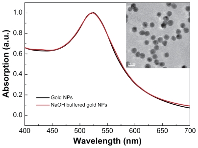
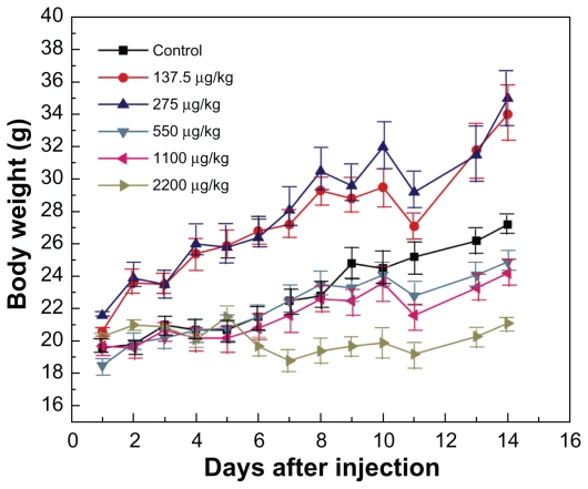
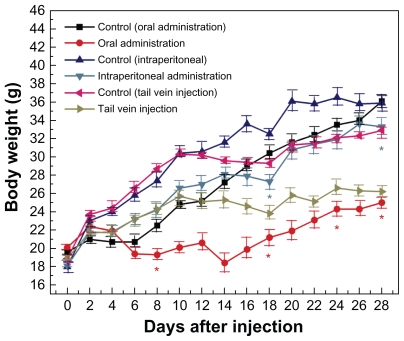
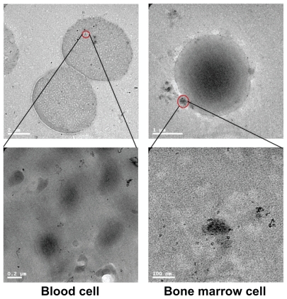
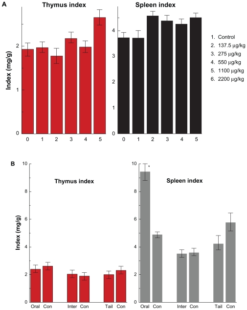
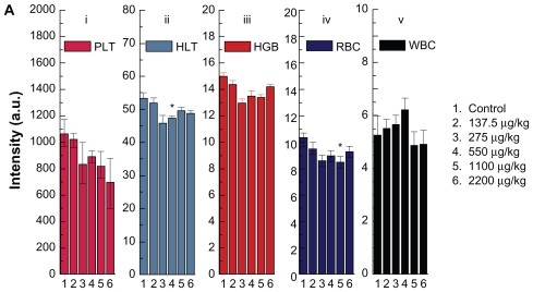
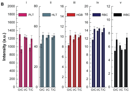
Gold nanoparticles have potential applications in biomedicine, but one of the important concerns is about their safety. Most toxicology data are derived from in vitro studies and may not reflect in vivo responses. Here, an animal toxicity study of 13.5 nm gold nanoparticles in mice is presented. Animal survival, weight, hematology, morphology, and organ index are characterized at different concentrations (137.5-2200 μg/kg) over 14-28 days. The results show that low concentrations of gold nanoparticles do not cause an obvious decrease in body weight or appreciable toxicity, even after their breakdown in vivo. High concentrations of gold nanoparticles induced decreases in body weight, red blood cells, and hematocrit. It was also found that gold nanoparticles administered orally caused significant decreases in body weight, spleen index, and red blood cells. Of the three administration routes, the oral and intraperitoneal routes showed the highest toxicity, and the tail vein injection showed the lowest toxicity. Combining the results of all of these studies, we suggest that targeted gold nanopartices by tail vein injection may be suitable for enhancement of radiotherapy, photothermal therapy, and related medical diagnostic procedures.

Keywords: gold nanoparticles; in vivo; toxicity.


Figures

Figure 1
Figure 1
Size and morphology of gold nanoparticles. The ultraviolet spectra of gold nanoparticles and transmission electron microscopic analysis (figure inset). Note: Gold nanoparticles were synthesized following the procedure described by Turkevich et al.
Figure 2
Figure 2
Body weight changes for mice treated with gold nanoparticles at doses of 137.5–2200 μg/kg. Note: Body weight was measured every two days. Each point represents the mean ± standard deviation of six mice. Data were analyzed by Student’s t-test and the differences between the doses and control group for each organ are not significant (P > 5%).
Figure 3
Figure 3
Body weight changes in mice treated with gold nanoparticles 1100 μg/kg by using three administration routes, ie, oral, intraperitoneal, and tail vein injection. Note: Each point represents mean ± standard deviation. Data were analyzed by Student’s t-test. *Represents significant difference from the control group (P < 0.05).
Figure 4
Figure 4
Transmission electron microscopy figures for gold nanoparticles in bone marrow and blood cells 14 days after oral administration at 2200 μg/kg.
Figure 5
Figure 5
Thymus and spleen indices of mice after oral administration by different doses (137.5–2200 μg/kg) (A) after 14 days and the different injection routes at the dose of 1100 μg/kg (B) after 28 days. Note: All values are reported as means ± standard deviation. Data were analyzed by Student’s t- test. *Represents significant difference from the control group (P < 0.05). Abbreviations: ORAL, oral administration; INTER, intraperitoneal injection; TAIL, tail vein injection.
Figure 6a
Figure 6a
Hematology results from mice treated with gold nanoparticles and the control group after 14 days oral administration by different doses (137.5–2200 μg/kg). Note: Bars represent mean ± standard deviation. Data were analyzed by Student’s t-test. *Represents significant difference from the control group (P < 0.05). Abbreviations: PLT, platelets; HCT, hematocrit; HGB, hemoglobin; RBC, red blood cells; WBC, white blood cells.
Figure 6b
Figure 6b
Hematology results from mice treated with gold nanoparticles and control groups after 28 days oral administration, intraperitoneal injection, and tail vein injection at the dose of 1100 μg/kg. Note: Bars represent mean ± standard deviation. Data were analyzed by Student’s t-test. *Represents significant difference from the control group (P < 0.05). Abbreviations: PLT, platelets; HCT, hematocrit; HGB, hemoglobin; RBC, red blood cells; WBC, white blood cells; O/C, oral administration control; I/C, intraperitoneal injection/control; T/C, tail vein injection/control.

References

    1. Danieland MC, Astruc D. Gold nanoparticles: Assembly, supramolecular chemistry, quantum-size-related properties, and applications toward biology, catalysis, and nanotechnology. Chem Rev. 2004;104:293–346. - PubMed
    1. Eustis S, El-Sayed MA. Why gold nanoparticles are more precious than pretty gold: Noble metal surface plasmon resonance and its enhancement of the radiative and nonradiative properties of nanocrystals of different shapes. Chem Soc Rev. 2006;35:209–217. - PubMed
    1. Hu M, Chen J, Li ZY, et al. Gold nanostructures: Engineering their plasmonic properties for biomedical applications. Chem Soc Rev. 2006;35:1084–94. - PubMed
    1. Anker JN, Hall WP, Lyandres O, Shah NC, Zhao J, van Duyne RP. Biosensing with plasmonic nanosensors. Nat Mater. 2008;7:442–453. - PubMed
    1. Sokolov K, Follen M, Aaron J, et al. Real-time vital optical imaging of precancer using anti-epidermal growth factor receptor anti-bodies conjugated to gold nanoparticles. Cancer Res. 2003;63:1999–2004. - PubMed

Publication types

MeSH terms