

Multiple neural tube defects in the same patient with no neurological deficit
- PMID: 21042511
- PMCID: PMC2964786
- DOI: 10.4103/1817-1745.66677
Multiple neural tube defects in the same patient with no neurological deficit
Abstract
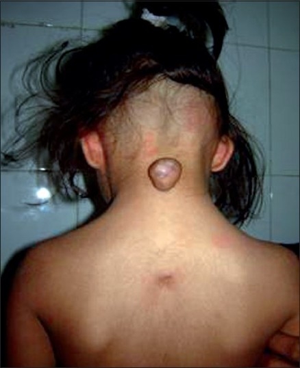
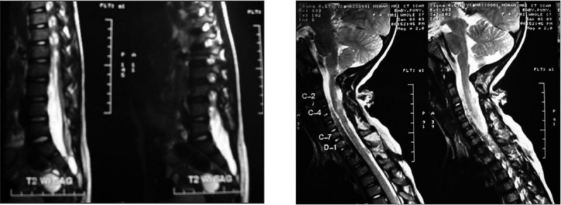
Congenital deformities involving the coverings of the nervous system are called neural tube defects (NTDs). NTD can be classified as neurulation defects, which occur by stage 12, and postneurulation defects. Cervical meningocele and myelomeningocele are rare spinal dysraphic lesions. Unlike lumbosacral dysraphic lesions, there is often no neurologic deficits and thus the subtle features of cervical cord tethering may be overlooked on imaging. The presence of meningomyelocele and/or encephaloceles at multiple (two or more) sites along the vertebral axis is a very rare event occurring in <1% of cases. Less than 10 cases have been described in the published literature. We are reporting a case of multiple NTD in same patient with no neurological deficit.
Keywords: Dermal sinus; meningomyelocele; neral tube defect; tethered cord.
Conflict of interest statement
Figures
References
-
- Lemire RJ. Neural tube defects. JAMA. 1988;259:558–62. - PubMed
-
- Ahmad FU, Dwarakanath S, Sharma BS, Mahapatra AK. Multiple neural tube defects: A clinical series of seven cases and their embryological basis. Pediatr Neurosurg. 2008;44:280–7. - PubMed
-
- Fisher RG, Uihlein A, Keith HM. Spina bifida and cranium bifidum: study of 530 cases. Proc Staff Meet Mayo Clin. 1952;27:33–8. - PubMed
-
- Barson AJ. Spina bifida: The significance of the level and extent of the defect to the morphogenesis. Dev Med Child Neurol. 1970;12:129–44. - PubMed
