. 2010 Oct 15:16:2092-108.

Neuroinflammation in advanced canine glaucoma

Affiliations

Neuroinflammation in advanced canine glaucoma

Bing Jiang et al. Mol Vis. .

Abstract

Purpose: The pathophysiological events that occur in advanced glaucoma are not well characterized. The principal purpose of this study is to characterize the gene expression changes that occur in advanced glaucoma.

Methods: Retinal RNA was obtained from canine eyes with advanced glaucoma as well as from healthy eyes. Global gene expression patterns were determined using oligonucleotide microarrays and confirmed by real-time PCR. The presence of tumor necrosis factor (TNF) and its receptors was evaluated by immunolabeling. Finally, we evaluated the presence of serum autoantibodies directed against retinal epitopes using western blot analyses.

Results: We identified over 500 genes with statistically significant changes in expression level in the glaucomatous retina. Decreased expression levels were detected for large number of functional groups, including synapse and synaptic transmission, cell adhesion, and calcium metabolism. Many of the molecules with decreased expression levels have been previously shown to be components of retinal ganglion cells. Genes with elevated expression in glaucoma are largely associated with inflammation, such as antigen presentation, protein degradation, and innate immunity. In contrast, expression of many other pro-inflammatory genes, such as interferons or interleukins, was not detected at abnormal levels.

Conclusions: This study characterizes the molecular events that occur in the canine retina with advanced glaucoma. Our data suggest that in the dog this stage of the disease is accompanied by pronounced retinal neuroinflammation.


Figures

Figure 1
Figure 1
Morphology of the canine retina. Morphology of the peripheral retina in a healthy dog (A) and dog with advanced glaucoma (B). Glaucomatous changes include dramatic loss of cells in the retinal nerve fiber layer, ganglion cell layer and inner nuclear layer compared to healthy eyes. In the glaucomatous retina GFAP can readily be detected in retinal glial cells (arrows) and indicates extensive gliosis in the nerve fiber layer. GFAP can also be detected in the NFL of normal eyes when extended periods of color development are used. (NFL-nerve fiber layer, RGC-retinal ganglion cell layer, IPL-inner plexiform layer, INL-inner nuclear layer, OPL-outer plexiform layer, ONL-outer nuclear layer, OS-Photoreceptor cell outer segments).
Figure 2
Figure 2
Volcano plot of the gene expression changes in the canine glaucomatous retina. Negative expression changes denote transcripts detected at reduced levels in glaucomatous eyes. This plot also represents all expressed genes, including those with less than twofold expression changes. Vertical bars represent a twofold expression change. Probability values were derived by Student’s t-test.
Figure 3
Figure 3
Verification of selected expression changes by quantitative PCR. Elevated expression could be confirmed for colony stimulating factor 1 receptor (CSF1R), glia maturation factor gamma (GMFG), neuronal cell adhesion molecule (NRCAM), neurotrophin3 (NTR3), Calgizzarin (S100A1), TNF receptor 1 (TNFRSF1A), and TNF receptor 14 (TNFRSF14). Error bars signify standard error.
Figure 4
Figure 4
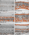
Digitalized images of immunohistochemistry based protein expression, which were used for quantification purposes. Increased TNF alpha expression was detected in glaucomatous eyes (B), predominantly in the nerve fiber layer, when compared to the control eyes (A). TNF alpha receptor 1 protein expression had similar appearance in control and glaucomatous eyes (C, D). TNF alpha receptor 2 protein expression was higher in glaucomatous eyes (F) when compared to control eyes (E).
Figure 5
Figure 5
Quantitative analysis of TNF, TNFR1, and TNFR2 expression in the glaucomatous retina. Quantitative analysis of TNFa (A), TNF receptor 1 (B), and TNF receptor 2 (C) immune reactivity in the central and peripheral retina of dogs with and without glaucoma. Statistical analyses reveal significantly higher TNF-alpha (A) and TNF-alpha receptor 2 (C) expression in glaucomatous eyes, when compared to control eyes. There was no significant difference in TNF alpha receptor 2 expression between control and glaucomatous eyes (B).
Figure 6
Figure 6
Immunohistochemical detection of CD3. CD3 positive cells are not apparent in the canine glaucomatous retina (left). In contrast, CD3 positive cells can easily be detected in optic nerve sections of a canine patient with optic neuritis (positive control). Note labeled cells in the perivascular space of the optic nerve (arrows). ONL-outer nuclear layer, INL-inner nuclear layer, NFL – nerve fiber layer.
Figure 7
Figure 7
Western blotting analysis of autoantibodies present in sera of dogs with glaucoma and in sera of control healthy dog sera. Sera from dogs with glaucoma are labeled from 1 to 7; control dog sera are labeled as “control dog sera,” Lanes R- recoverin, E – enolase, C - crystalline-μ are immunostained with antibodies specifically directed against these molecules. N – negative control, M – molecular weight markers.

Similar articles

Cited by

References

    1. Grozdanic SD, Matic M, Betts DM, Sakaguchi DS, Kardon RH. Recovery of canine retina and optic nerve function after acute elevation of intraocular pressure: implications for canine glaucoma treatment. Vet Ophthalmol. 2007;10:101–7. - PubMed
    1. Grozdanic S, Kecova H, Harper MM, Nilaweera WU, Kuehn MH, Kardon RH. Functional and structural changes in a canine model of hereditary primary angle-closure glaucoma. Invest Ophthalmol Vis Sci. 2010;51:255–63. - PMC - PubMed
    1. Ahmed F, Brown KM, Stephan DA, Morrison JC, Johnson EC, Tomarev SI. Microarray analysis of changes in mRNA levels in the rat retina after experimental elevation of intraocular pressure. Invest Ophthalmol Vis Sci. 2004;45:1247–58. - PubMed
    1. Naskar R, Thanos S. Retinal gene profiling in a hereditary rodent model of elevated intraocular pressure. Mol Vis. 2006;12:1199–210. - PubMed
    1. Steele MR, Inman DM, Calkins DJ, Horner PJ, Vetter ML. Microarray analysis of retinal gene expression in the DBA/2J model of glaucoma. Invest Ophthalmol Vis Sci. 2006;47:977–85. - PubMed

Publication types

MeSH terms