. 2010 Oct 22;5(10):e13563.
doi: 10.1371/journal.pone.0013563.

Familial glucocorticoid receptor haploinsufficiency by non-sense mediated mRNA decay, adrenal hyperplasia and apparent mineralocorticoid excess

Affiliations

Familial glucocorticoid receptor haploinsufficiency by non-sense mediated mRNA decay, adrenal hyperplasia and apparent mineralocorticoid excess

Jérôme Bouligand et al. PLoS One. .

Abstract

Primary glucocorticoid resistance (OMIM 138040) is a rare hereditary disease that causes a generalized partial insensitivity to glucocorticoid action, due to genetic alterations of the glucocorticoid receptor (GR). Investigation of adrenal incidentalomas led to the discovery of a family (eight affected individuals spanning three generations), prone to cortisol resistance, bilateral adrenal hyperplasia, arterial hypertension and hypokalemia. This phenotype exacerbated over time, cosegregates with the first heterozygous nonsense mutation p.R469[R,X] reported to date for the GR, replacing an arginine (CGA) by a stop (TGA) at amino-acid 469 in the second zinc finger of the DNA-binding domain of the receptor. In vitro, this mutation leads to a truncated 50-kDa GR lacking hormone and DNA binding capacity, devoid of hormone-dependent nuclear translocation and transactivation properties. In the proband's fibroblasts, we provided evidence for the lack of expression of the defective allele in vivo. The absence of detectable mutated GR mRNA was accompanied by a 50% reduction in wild type GR transcript and protein. This reduced GR expression leads to a significantly below-normal induction of glucocorticoid-induced target genes, FKBP5 in fibroblasts. We demonstrated that the molecular mechanisms of glucocorticoid signaling dysfunction involved GR haploinsufficiency due to the selective degradation of the mutated GR transcript through a nonsense-mediated mRNA Decay that was experimentally validated on emetine-treated propositus' fibroblasts. GR haploinsufficiency leads to hypertension due to illicit occupation of renal mineralocorticoid receptor by elevated cortisol rather than to increased mineralocorticoid production reported in primary glucocorticoid resistance. Indeed, apparent mineralocorticoid excess was demonstrated by a decrease in urinary tetrahydrocortisone-tetrahydrocortisol ratio in affected patients, revealing reduced glucocorticoid degradation by renal activity of the 11β-hydroxysteroid dehydrogenase type 2, a GR regulated gene. We propose thus that GR haploinsufficiency compromises glucocorticoid sensitivity and may represent a novel genetic cause of subclinical hypercortisolism, incidentally revealed bilateral adrenal hyperplasia and mineralocorticoid-independent hypertension.


Conflict of interest statement

Competing Interests: The authors have declared that no competing interests exist.

Figures

Figure 1
Figure 1. A family with glucocorticoid resistance.
A) Structure of the pedigree. Three generations (I, II and III) of the kindred are represented. Individuals carrying the heterozygous R469X mutation are shown in black. Black arrow indicates the proband. B) Phenotype of the propositus. C) Bilateral adrenal hyperplasia was readily visible by computerized tomography (CT) in three affected individuals belonging to three generations (I.2, the mother of the propositus; II.3, the propositus; and III.2, his daughter, white arrows indicate adrenal glands).
Figure 2
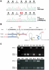
Figure 2. Identification of the GR R469X mutation in the kindred.
A) Identification of the heterozygous 1405C>T transition. Sequencing of exon 4 in genomic DNA of individual II.1 confirmed the existence of the normal GR coding region, whereas the corresponding sequence of the proband DNA indicated that patient II.3 was heterozygous for a single C>T nucleotide change at position 1405, converting the amino acid arginine (R) into a premature stop codon (X) at position 469 of GR in all affected individuals. B) Genomic organization of the human GR and functional domains of the wildtype GRα. The hGR gene is composed of 10 exons, the two last two of which exon 9α and exon 9β are alternatively spliced. The C>T substitution at position 1405 in exon 4 results in a premature termination of translation and gives rise to a 468-amino-acid truncated GR mutant which lacks the C-terminal region of the receptor including part of the DNA-binding domain (DBD) and the ligand-binding domain (LBD). C) The 1405C>T substitution abrogated the Bsp119I restriction site in exon 4 of GR, thus allowing rapid identification of the heterozygous mutation. PCR-amplified exon 4 fragments from all individuals in the kindred were digested with Bsp119I and loaded on agarose gel: (upper panel individuals I.2 to II.7, lower panel individuals III.1 to III.6). The presence of a 286-bp fragment resistant to Bsp119I digestion confirmed the C>T substitution.
Figure 3
Figure 3. In vitro characterization of the GR mutant.
A) Western blot analysis of GR. Four micrograms of protein obtained from homogenates of HEK293 cells transiently transfected with WT hGRα (WT) and hGRα-R469X mutant were processed for immunoblotting with an anti-GR antibody. Note the presence of a specific 90-kDa band for WT GR and a 50-kDa band for hGRα-R469X. B) Protein expression of in vitro-translated [35S]-labeled WT hGRα and hGRα-R469X separated by 10% SDS-PAGE. GR migrated as a major 90-kDa form whereas the GR mutant, as expected, had a lower molecular mass of approximately 50 kDa. C) Binding of in vitro-translated WT hGRα and hGRα-R469X to GRE consensus sequence by gel retardation assay. Specific GR-radiolabeled GRE complexes (arrow) were detected in the absence of unlabeled competitor (-), which were abolished in the presence of 50 ng unlabeled probe (50). As expected, the GR mutant was unable to bind DNA. P: free probe. D) Intracellular trafficking of WT hGRα and hGRα-R469X in transfected COS7 cells by Immunocytochemistry. Cells were counterstained with DAPI in blue. WT GR translocates from the cytoplasm to the nucleus after 5 min incubation with 1 mM DXM whereas GR mutant remains exclusively in the cytoplasmic compartment either in the absence or presence of DXM. E) Transcriptional activity of the WT hGRαand the truncated hGRα-R469X mutant. HEK 293 cells were transfected using Lipofectamin 2000 with either WT hGRα or hGRα-R469X together with the glucocorticoid-responsive reporter gene pGL3-GRE2-TATA-Luc and pSV.β-Gal plasmids. Following transfection, cells were exposed to 100 nM DXM for 24 h. Results (Luc/β-gal activity) are expressed as the percentage of relative transcriptional activity of WT GR arbitrarily set at 100% with 100 nM DXM. Results are means ± SD of at least 3 independent determinations.
Figure 4
Figure 4. Evidence of GR haploinsufficiency in the proband's fibroblasts due to nonsense-mediated mRNA Decay.
A) Demonstration of GR haploinsufficiency in the fibroblasts of the propositus (II.3). Sequencing of exon 4 genomic DNA prepared from the patient's fibroblasts confirmed the presence of the heterozygous C>T substitution as observed in lymphocyte genomic DNA (see Supplemental Fig. S1A SI lower panel). In contrast, direct sequencing of the cDNA (see specific primers in Supplemental Table S1 SI) prepared from fibroblast RNA revealed only the wildtype C allele. The absence of mutated GR transcripts (upper panel) in the patient's fibroblasts is consistent with nonsense-mediated mRNA Decay, a cellular mechanism that prevents translation of mutated mRNA bearing a premature termination codon. When fibroblasts were treated for 6 h with 100 µg/ml emetine (lower panel) or for 2 h with 20 µg/ml cycloheximide (not shown), the expression of the defective allele was restored as shown by direct sequencing of the corresponding cDNA fragment. B) Increase in GR mRNA expression in fibroblasts of patient II.3 after exposure to two inhibitors of nonsense-mediated mRNA Decay, cycloheximide (20 µg/ml for 2 h) or emetine (100 µg/ml for 6 h). Relative expression of GR, beta-actin ou 18S RNA was measured by using quantitative real-time RT-PCR. Results are means ± SEM of 4 determinations and expressed as fold induction relative to untreated cells. (* P<0.05 Kruskal Wallis followed by Dunn's post test and Mann Whitney test). C) Reduction of GR mRNA expression in fibroblasts of patient II.3 compared with two controls C1 and C2. The expression of mRNA was measured by using quantitative real-time RT-PCR. Results are expressed as attomol/fmol of 18S and are means ± SEM of 3 independent determinations (*** P<0.001 Mann Whitney test). D) Reduction in specific [3H]-DXM binding sites. Fibroblasts pre-incubated in steroid-free medium for 24 h, were exposed to 50 nM [3H]-DXM in the absence or presence of a 500-fold excess of unlabeled DEX for 1 h at 37°C. Radioactivity was measured and specific binding was calculated. Data are means ± SEM of 3 independent determinations performed in triplicate. The estimated GR density in the propositus' fibroblasts was 3×10⁴ sites per cell (*** P<0.001 vs controls). E) Western blot analysis of GR. Thirty micrograms of protein from fibroblast homogenates of controls (C1 and C2) and patient II.3 were processed for immunoblotting with anti-GR (upper panel) and anti-b actin (lower panel). Note the presence of a specific 90-kDa GR in controls and an approximately 50% reduction in WT GR expression in patient II.3 whereas the 50-kDa band corresponding to the truncated hGRa-R469X mutant was not detected. Quantitative analysis of GR signals normalized to b-actin loading was performed using QuantityOne software (Biorad). Results are means ± SD of at least 3 independent analyses (* P<0.05 Mann Whitney test). F) Altered glucocorticoid-inducible gene expression in the patient's fibroblasts. Fibroblasts from controls (C1 and C2) and from patient II.3 were starved for 24 h in steroid-free medium and then exposed to 100 nM DXM for 6 h. Relative levels of FKBP5 transcripts were determined by quantitative real-time RT-PCR analysis. Results are expressed as attomol/fmol 18S and are means ± SEM of 6 independent determinations (** P<0.01, Kruskal Wallis followed by Dunn's post test).

References

    1. Lu NZ, Wardell SE, Burnstein KL, Defranco D, Fuller PJ, et al. International Union of Pharmacology. LXV. The pharmacology and classification of the nuclear receptor superfamily: glucocorticoid, mineralocorticoid, progesterone, and androgen receptors. Pharmacol Rev. 2006;58:782–797. - PubMed
    1. Lu NZ, Cidlowski JA. The origin and functions of multiple human glucocorticoid receptor isoforms. Ann N Y Acad Sci. 2004;1024:102–123. - PubMed
    1. Rhen T, Cidlowski JA. Antiinflammatory action of glucocorticoids–new mechanisms for old drugs. N Engl J Med. 2005;353:1711–1723. - PubMed
    1. Gross KL, Cidlowski JA. Tissue-specific glucocorticoid action: a family affair. Trends Endocrinol Metab. 2008;19:331–339. - PMC - PubMed
    1. Viengchareun S, Le Menuet D, Martinerie L, Munier M, Pascual-Le Tallec L, et al. The mineralocorticoid receptor: insights into its molecular and (patho)physiological biology. Nucl Recept Signal. 2007;5:e012. - PMC - PubMed

Publication types