

Molecular neurobiology of lead (Pb(2+)): effects on synaptic function
- PMID: 21042954
- PMCID: PMC3076195
- DOI: 10.1007/s12035-010-8146-0
Molecular neurobiology of lead (Pb(2+)): effects on synaptic function
Abstract
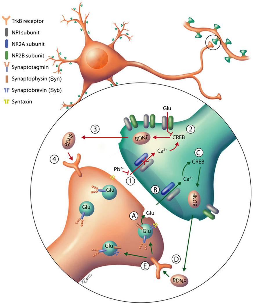
Lead (Pb(2+)) is a ubiquitous environmental neurotoxicant that continues to threaten public health on a global scale. Epidemiological studies have demonstrated detrimental effects of Pb(2+) on childhood IQ at very low levels of exposure. Recently, a mechanistic understanding of how Pb(2+) affects brain development has begun to emerge. The cognitive effects of Pb(2+) exposure are believed to be mediated through its selective inhibition of the N-methyl-D: -aspartate receptor (NMDAR). Studies in animal models of developmental Pb(2+) exposure exhibit altered NMDAR subunit ontogeny and disruption of NMDAR-dependent intracellular signaling. Additional studies have reported that Pb(2+) exposure inhibits presynaptic calcium (Ca(2+)) channels and affects presynaptic neurotransmission, but a mechanistic link between presynaptic and postsynaptic effects has been missing. Recent work has suggested that the presynaptic and postsynaptic effects of Pb(2+) exposure are both due to inhibition of the NMDAR by Pb(2+), and that the presynaptic effects of Pb(2+) may be mediated by disruption of NMDAR activity-dependent signaling of brain-derived neurotrophic factor (BDNF). These findings provide the basis for the first working model to describe the effects of Pb(2+) exposure on synaptic function. Here, we review the neurotoxic effects of Pb(2+) exposure and discuss the known effects of Pb(2+) exposure in light of these recent findings.
Figures
Similar articles
-
Neurotoxicity of lead. Hypothetical molecular mechanisms of synaptic function disorders.Neurol Neurochir Pol. 2012 Nov-Dec;46(6):569-78. doi: 10.5114/ninp.2012.31607. Neurol Neurochir Pol. 2012. PMID: 23319225 Review.
-
Lead exposure during synaptogenesis alters vesicular proteins and impairs vesicular release: potential role of NMDA receptor-dependent BDNF signaling.Toxicol Sci. 2010 Jul;116(1):249-63. doi: 10.1093/toxsci/kfq111. Epub 2010 Apr 7. Toxicol Sci. 2010. PMID: 20375082 Free PMC article.
-
Enhanced nitric oxide production during lead (Pb²⁺) exposure recovers protein expression but not presynaptic localization of synaptic proteins in developing hippocampal neurons.Brain Res. 2012 Feb 23;1439:88-95. doi: 10.1016/j.brainres.2011.12.037. Epub 2011 Dec 29. Brain Res. 2012. PMID: 22265330 Free PMC article.
-
Lead exposure during synaptogenesis alters NMDA receptor targeting via NMDA receptor inhibition.Neurotoxicology. 2011 Mar;32(2):281-9. doi: 10.1016/j.neuro.2010.12.013. Epub 2010 Dec 28. Neurotoxicology. 2011. PMID: 21192972 Free PMC article.
-
Lead neurotoxicity: from exposure to molecular effects.Brain Res Brain Res Rev. 2005 Nov;49(3):529-54. doi: 10.1016/j.brainresrev.2005.02.004. Epub 2005 Mar 31. Brain Res Brain Res Rev. 2005. PMID: 16269318 Review.
Cited by
-
Presynaptic mechanisms of lead neurotoxicity: effects on vesicular release, vesicle clustering and mitochondria number.PLoS One. 2015 May 26;10(5):e0127461. doi: 10.1371/journal.pone.0127461. eCollection 2015. PLoS One. 2015. PMID: 26011056 Free PMC article.
-
Genome-Wide Analyses of Metal Responsive Genes in Caenorhabditis elegans.Front Genet. 2012 Apr 10;3:52. doi: 10.3389/fgene.2012.00052. eCollection 2012. Front Genet. 2012. PMID: 22514555 Free PMC article.
-
Therapeutic role of garlic and vitamins C and E against toxicity induced by lead on various organs.Environ Sci Pollut Res Int. 2020 Mar;27(9):8953-8964. doi: 10.1007/s11356-020-07654-2. Epub 2020 Feb 8. Environ Sci Pollut Res Int. 2020. PMID: 32036533 Review.
-
Long-term probiotic intervention mitigates memory dysfunction through a novel H3K27me3-based mechanism in lead-exposed rats.Transl Psychiatry. 2020 Jan 22;10(1):25. doi: 10.1038/s41398-020-0719-8. Transl Psychiatry. 2020. PMID: 32066679 Free PMC article.
-
The Effect of Lead Exposure on Autism Development.Int J Mol Sci. 2021 Feb 6;22(4):1637. doi: 10.3390/ijms22041637. Int J Mol Sci. 2021. PMID: 33561959 Free PMC article. Review.
References
-
- Byers RK, Lord EE. Late effects of lead poisoning on mental development. Am J Dis Child. 1943;66:471–494.
-
- Needleman HL, Gatsonis CA. Low-level lead exposure and the IQ of children. A meta-analysis of modern studies. JAMA. 1990;263:673–678. - PubMed
-
- Lanphear BP, Hornung R, Khoury J, Yolton K, Baghurst P, Bellinger DC, Canfield RL, Dietrich KN, Bornschein R, Greene T, Rothenberg SJ, Needleman HL, Schnaas L, Wasserman G, Graziano J, Roberts R. Low-level environmental lead exposure and children’s intellectual function: an international pooled analysis. Environ Health Perspect. 2005;113:894–899. - PMC - PubMed
Publication types
MeSH terms
Substances
Grants and funding
LinkOut - more resources
Full Text Sources
Miscellaneous
