. 2011 Dec;5(4):531-45.
doi: 10.3109/17435390.2010.530004. Epub 2010 Nov 3.

Mast cells contribute to altered vascular reactivity and ischemia-reperfusion injury following cerium oxide nanoparticle instillation

Affiliations

Mast cells contribute to altered vascular reactivity and ischemia-reperfusion injury following cerium oxide nanoparticle instillation

Christopher J Wingard et al. Nanotoxicology. 2011 Dec.

Abstract

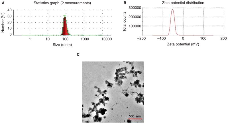
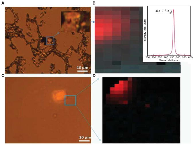
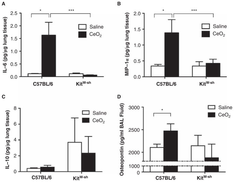
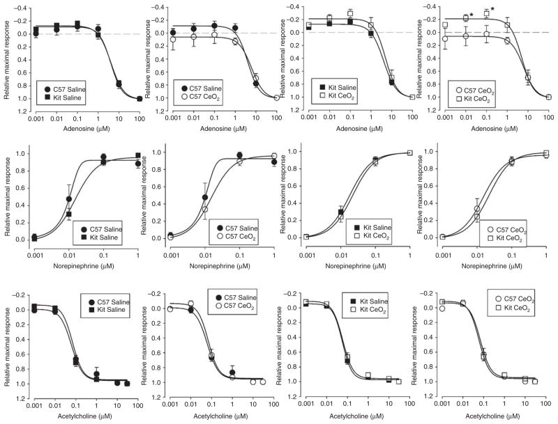
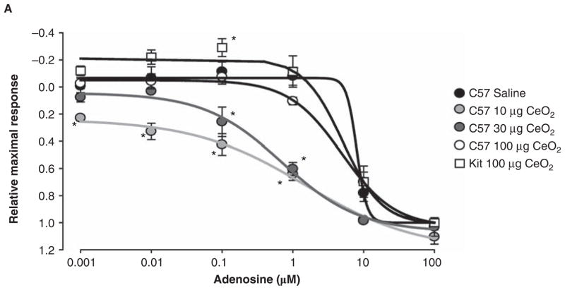
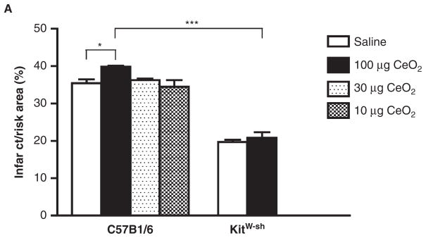
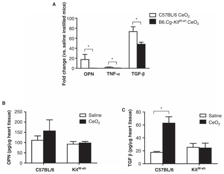
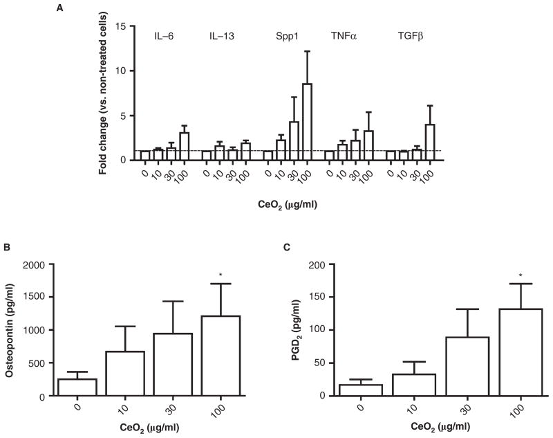
Cerium oxide (CeO₂) represents an important nanomaterial with wide ranging applications. However, little is known regarding how CeO₂ exposure may influence pulmonary or systemic inflammation. Furthermore, how mast cells would influence inflammatory responses to a nanoparticle exposure is unknown. We thus compared pulmonary and cardiovascular responses between C57BL/6 and B6.Cg-Kit(W-sh) mast cell deficient mice following CeO₂ nanoparticle instillation. C57BL/6 mice instilled with CeO₂ exhibited mild pulmonary inflammation. However, B6.Cg-Kit(W-sh) mice did not display a similar degree of inflammation following CeO₂ instillation. Moreover, C57BL/6 mice instilled with CeO₂ exhibited altered aortic vascular responses to adenosine and an increase in myocardial ischemia/reperfusion injury which was absent in B6.Cg-Kit(W-sh) mice. In vitro CeO₂ exposure resulted in increased production of PGD₂, TNF-α, IL-6 and osteopontin by cultured mast cells. These findings demonstrate that CeO₂ nanoparticles activate mast cells contributing to pulmonary inflammation, impairment of vascular relaxation and exacerbation of myocardial ischemia/reperfusion injury.


Conflict of interest statement

Declaration of interest: This work was supported by National Institute of Environmental Health Sciences (NIEHS) RO1ES016246 (CJW) and NIH RO1ES019311 (JMB). The authors declare they have no competing financial interests. The authors report no conflict of interest. The authors alone are responsible for the content and writing of the paper.

Figures

Figure 1
Figure 1
Characterization of CeO2 nanoparticles. (A) Hydrodynamic size distribution of CeO2 suspension obtained by two independent measurements, showing an average size of 90 ± 20 nm (shown in red). The standard deviations are shown in green. (B) Zeta potential of CeO2 suspension, indicating an average value of −52.7 mV. (C) TEM image of dehydrated CeO2 suspension. A solution of 1 μg/μl CeO2 was used for characterization.
Figure 2
Figure 2
Raman spectroscopy of CeO2 nanoparticles in mouse lung tissue. (A) An optical image of a CeO2-instilled C57BL/6 mouse lung section showing black colored CeO2 at the surface. (B) The corresponding Raman image of the mouse lung section shown in (A). The red colored region is a map of the characteristic Raman peak (shown in the inset ~ 464 cm−1) for CeO2 nanoparticles. (C) Optical image of CeO2 instilled mouse lung section showing CeO2 embedded at ~ 4 μm depth. (D) The corresponding Raman map of the 464 cm−1 peak for the section shown in (C).
Figure 3
Figure 3
Expression of inflammatory mediators in lung tissue and BALF in C57BL/6 and B6.Cg-KitW-sh mice 24 hours after instillation of 100 μg CeO2. (A) IL-6, (B) MIP-1α, (C) IL-10 protein levels measured in lung tissue 24 h following instillation of CeO2 in C57BL/6 and B6.Cg-KitW-sh mice. (D) Osteopontin protein levels measured in BALF of saline or CeO2 instilled C57BL/6 and B6.Cg-KitW-sh mice. n = 7–9 mice/group. p ≤ 0.05.
Figure 4
Figure 4
Effect of CeO2 instillation on the contraction and relaxation of isolated thoracic aorta from C57BL/6 or B6.Cg-KitW-sh mice. (A) Adenosine concentration response profiles; (B) norepinephrine concentration response profiles and (C) acetylcholine concentration response profiles. Aortic rings were prestimulated with 1 μM phenylephrine. n = 6–10 mice/group. p ≤ 0.05 versus C57BL/6.
Figure 5
Figure 5
Comparison of the adenosine dose-response of thoracic aortic ring segments from C57BL/6 mice exposed to 10, 30 and 100 μg of CeO2 and B6.Cg-KitW-sh mice exposed to 100 μg of CeO2. n = 6–10 mice/group. p ≤ 0.05 as compared to saline treated mice.
Figure 6
Figure 6
Comparison of the size of myocardial infarction in C57BL/6 exposed to 10, 30 and 100 μg of CeO2 and B6.Cg-KitW-sh mice exposed to 100 μg of CeO2. Twenty-four hours after instillation with either saline or CeO2, the LAD was ligated for 20 min and then reperfused for 2 h. n = 3–4 mice/group. p ≤ 0.05 ★★★ p ≤ 0.001.
Figure 7
Figure 7
Expression of osteopontin, TNF-α and TGF-β in non-infarcted heart tissue following CeO2 instillation. (A) mRNA levels of osteopontin, TNF-α and TGF-β in non-infarcted heart tissue following CeO2 instillation in C57BL/6 or B6.Cg-KitW-sh mice. (B) Osteopontin and (C) TGF-β protein levels in non-infarcted heart tissue following CeO2 instillation in C57BL/6 or B6.Cg-KitW-sh mice. n = 3–4 mice/group. p ≤ 0.05.
Figure 8
Figure 8
Effect of CeO2 on in vitro BMMC activation. (A) Fold change in mRNA expression of IL-6, IL-13, osteopontin (Spp1), TNF-α and TGF-β 2 hours following CeO2 treatment of BMMCs as compared to untreated cells. (B) Osteopontin protein levels measured in the supernatant of BMMCs exposed to CeO2 for 24 h as measured by ELISA. (C) PGD2 levels measured in supernatant of BMMCs exposed to CeO2 for 1 h as measured by EIA. n = 3–5 independent experiments. p ≤ 0.05.

Similar articles

Cited by

References

    1. Bhattacharya K, Farwell K, Huang M, Kempuraj D, Donelan J, Papaliodis D, Vasiadi M, Theoharides TC. Mast cell deficient W/Wv mice have lower serum IL-6 and less cardiac tissue necrosis than their normal littermates following myocardial ischemia-reperfusion. Int J Immunopathol Pharmacol. 2007;20:69–74. - PubMed
    1. Brook RD, Franklin B, Cascio W, Hong Y, Howard G, Lipsett M, Luepker R, Mittleman M, Samet J, Smith SC, Jr, Tager I. Air pollution and cardiovascular disease: A statement for health-care professionals from the Expert Panel on Population and Prevention Science of the American Heart Association. Circulation. 2004;109:2655–2671. - PubMed
    1. Brown JM, Swindle EJ, Kushnir-Sukhov NM, Holian A, Metcalfe DD. Silica-directed mast cell activation is enhanced by scavenger receptors. Am J Respir Cell Mol Biol. 2007;36:43–52. - PMC - PubMed
    1. Brown JM, Wilson TM, Metcalfe DD. The mast cell and allergic diseases: Role in pathogenesis and implications for therapy. Clin Exp Allergy. 2008;38:4–18. - PubMed
    1. Bulfone-Paus S, Paus R. Osteopontin as a new player in mast cell biology. Eur J Immunol. 2008;38:338–341. - PubMed

Publication types

MeSH terms