. 2010 Nov;32(10):1735-43.
doi: 10.1111/j.1460-9568.2010.07439.x. Epub 2010 Oct 7.

At the limbic-motor interface: disconnection of basolateral amygdala from nucleus accumbens core and shell reveals dissociable components of incentive motivation

Affiliations

At the limbic-motor interface: disconnection of basolateral amygdala from nucleus accumbens core and shell reveals dissociable components of incentive motivation

Michael W Shiflett et al. Eur J Neurosci. 2010 Nov.

Abstract

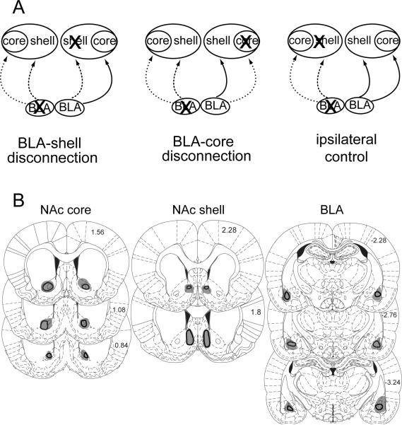
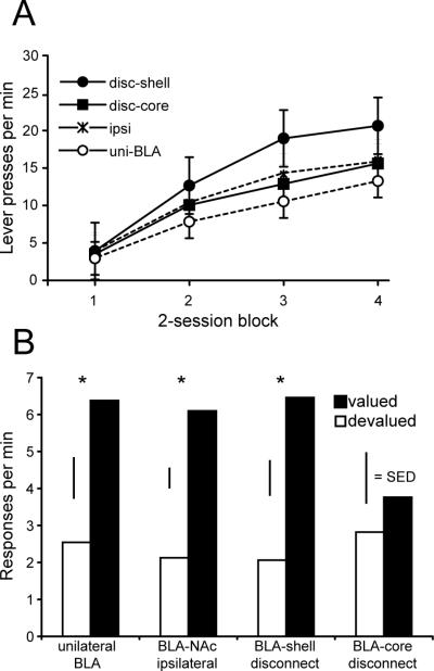
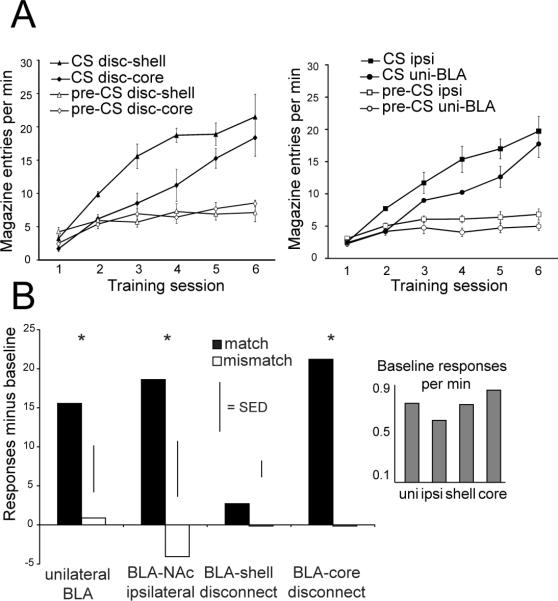
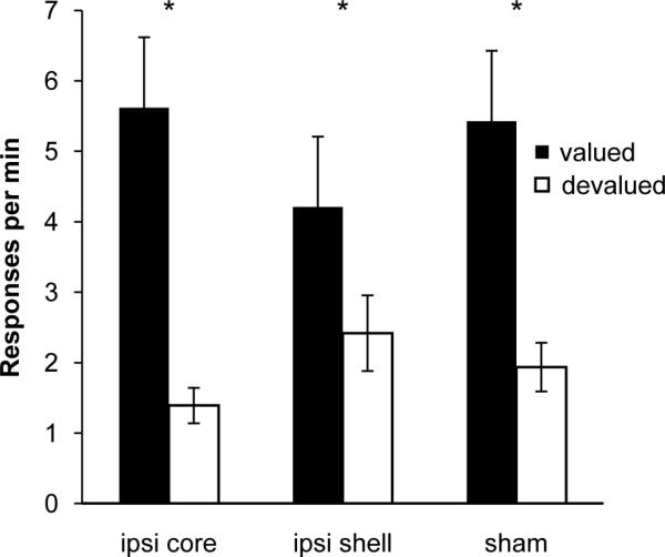
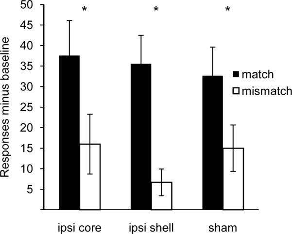
Although it has long been hypothesized that the nucleus accumbens (NAc) acts as an interface between limbic and motor regions, direct evidence for this modulatory role on behavior is lacking. Using a disconnection procedure in rats, we found that basolateral amygdala (BLA) input to the core and medial shell of the NAc separately mediate two distinct incentive processes controlling the performance of goal-directed instrumental actions, respectively: (i) the sensitivity of instrumental responding to changes in the experienced value of the goal or outcome, produced by specific satiety-induced outcome devaluation; and (ii) the effect of reward-related cues on action selection, observed in outcome-specific Pavlovian-instrumental transfer. These results reveal, therefore, that dissociable neural circuits involving BLA inputs to the NAc core and medial shell mediate distinct components of the incentive motivational processes controlling choice and decision-making in instrumental conditioning.


Figures

Figure 1
Figure 1. Histological results
A) Schematic of disconnection design. The ipsilateral control consisted of animals with either BLA-shell or BLA-core lesions B) Representation of the minimum (black outline) and maximum (gray shading) extent of tissue damage resulting from NAc core and shell lesions and BLA lesions. The left hemisphere represents lesions from control animals (ipsilateral BLA-NAc and unilateral BLA lesions). Lesions in the right hemisphere depict animals with disconnection of the core or shell from the BLA. Values indicate mm from Bregma. Reproduced from Paxinos and Watson.
Figure 2
Figure 2. BLA-Nac core disconnection impairs rats’ sensitivity to outcome value during instrumental performance
We trained rats over 8 sessions to bar press for food rewards and recorded the rate of responding across training. A) Rats from all groups acquired an instrumental response, as shown by an increase in response rates across training sessions. B) After one of the two instrumental outcomes was devalued by specific satiety, rats performed a choice test with both levers available for responding in extinction. Rats with unilateral BLA lesions (uni-BLA), ipsilateral NAc and BLA lesions (ipsi), and lesions that disconnect the NAc shell from the BLA bilaterally (disc-shell), made significantly fewer responses on the lever that, during training, resulted in delivery of the now devalued outcome. In contrast, disconnection of the NAc core from the BLA (disc-core) impaired rats’ ability to use outcome value to guide actions, as shown by the lack of a devaluation effect. * = non-devalued greater than devalued at P < 0.05, paired t-test with Bonferroni correction. Error bars = ± 1 SEM (panel A) or 1 standard error of the difference (SED) (panel B), a measure of within-subjects variability.
Figure 3
Figure 3. BLA-NAc shell disconnection impairs outcome-specific Pavlovian-instrumental transfer
A) We trained rats over 6 sessions to associate auditory stimuli with delivery of food pellets. During the session we measured the number of food cup entries during stimulus presentation (CS, filled symbols) and during an equivalent period prior to stimulus onset (pre-CS, open symbols). The graph depicts the number of food cup entries per min for each of the 6 training sessions. Data from the two disconnection groups are presented in the left panel and data for the two control groups in the right panel. We observed an increase in food cup entries across training sessions. In addition, rats with BLA-shell disconnection showed a greater number of head entries during stimulus presentations compared to the unilateral BLA lesion control condition. B) During the transfer test, two cues were presented while rats had access to both levers in extinction. Control rats (ipsi and uni-BLA) and rats with BLA-NAc core disconnection (disc-core) increased their rate of responding above baseline on the lever whose outcome during training matched the cue, compared to when cue and lever outcome were mismatched. In contrast, rats with BLA-NAc shell disconnection (disc-shell) showed no increase in instrumental performance above baseline during cue presentation. Inset depicts baseline responses per min measured during intervals between cue presentations. * = match greater than mismatch at P < 0.05, paired t-test with Bonferroni correction. Error bars = ± 1 SEM (panel A) or 1 SED (panel B).
Figure 4
Figure 4. Ipsilateral lesions do not disrupt choice performance following outcome devaluation
Rats respond at a greater rate on the lever that during training delivered the valued outcome compared to the devalued outcome. * = non-devalued greater than devalued at P < 0.05, paired t-test with Bonferroni correction. Error bars = ± 1 SEM
Figure 5
Figure 5. Ipsilateral BLA-NAc lesions do not impair performance during outcome-specific PIT
Rats respond at a greater rate on the lever whose outcome matches the cue (match) than on the mismatched lever regardless of lesion type. * = match greater than mismatch at P < 0.05, paired t-test with Bonferroni correction. Error bars = ± 1 SEM.

Similar articles

Cited by

References

    1. Ahn S, Phillips AG. Modulation by Central and Basolateral Amygdalar Nuclei of Dopaminergic Correlates of Feeding to Satiety in the Rat Nucleus Accumbens and Medial Prefrontal Cortex. J. Neurosci. 2002;22:10958–10965. - PMC - PubMed
    1. Alexander GE, Delong MR, Strick PL. Parallel organization of functionally segregated circuits linking basal ganglia and cortex. Annu Rev Neurosci. 1986;9:357–381. - PubMed
    1. Ambroggi F, Ishikawa A, Fields HL, Nicola SM. Basolateral Amygdala Neurons Facilitate Reward-Seeking Behavior by Exciting Nucleus Accumbens Neurons. Neuron. 2008;59:648–661. - PMC - PubMed
    1. Balleine B, Killcross S. Effects of ibotenic acid lesions of the nucleus accumbens on instrumental action. Behav Brain Res. 1994;65:181–193. - PubMed
    1. Balleine BW. Neural bases of food-seeking: affect, arousal and reward in corticostriatolimbic circuits. Physiol Behav. 2005;86:717–730. - PubMed

Publication types