. 2010 Dec;32(11):1912-20.
doi: 10.1111/j.1460-9568.2010.07467.x. Epub 2010 Oct 29.

Post-traumatic seizure susceptibility is attenuated by hypothermia therapy

Affiliations

Post-traumatic seizure susceptibility is attenuated by hypothermia therapy

Coleen M Atkins et al. Eur J Neurosci. 2010 Dec.

Abstract

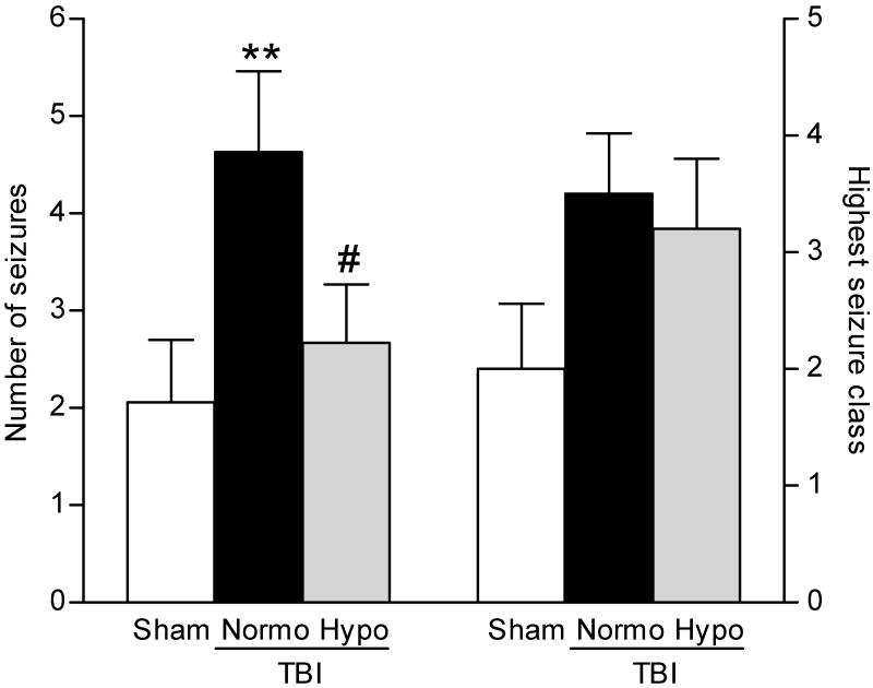
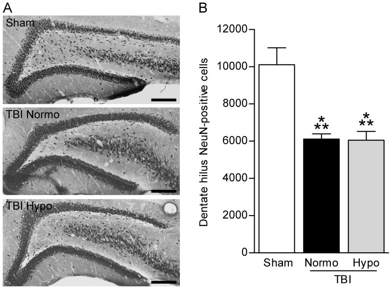
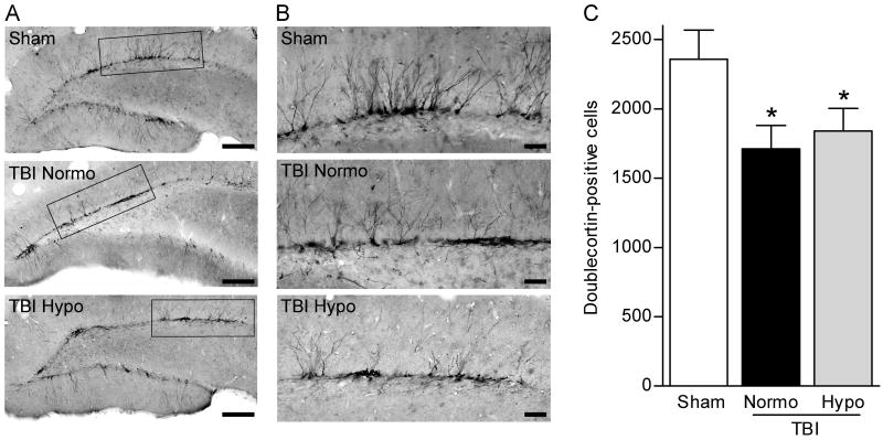
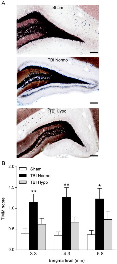
Traumatic brain injury (TBI) is a major risk factor for the subsequent development of epilepsy. Currently, chronic seizures after brain injury are often poorly controlled by available antiepileptic drugs. Hypothermia treatment, a modest reduction in brain temperature, reduces inflammation, activates pro-survival signaling pathways, and improves cognitive outcome after TBI. Given the well-known effect of therapeutic hypothermia to ameliorate pathological changes in the brain after TBI, we hypothesized that hypothermia therapy may attenuate the development of post-traumatic epilepsy and some of the pathomechanisms that underlie seizure formation. To test this hypothesis, adult male Sprague Dawley rats received moderate parasagittal fluid-percussion brain injury, and were then maintained at normothermic or moderate hypothermic temperatures for 4 h. At 12 weeks after recovery, seizure susceptibility was assessed by challenging the animals with pentylenetetrazole, a GABA(A) receptor antagonist. Pentylenetetrazole elicited a significant increase in seizure frequency in TBI normothermic animals as compared with sham surgery animals and this was significantly reduced in TBI hypothermic animals. Early hypothermia treatment did not rescue chronic dentate hilar neuronal loss nor did it improve loss of doublecortin-labeled cells in the dentate gyrus post-seizures. However, mossy fiber sprouting was significantly attenuated by hypothermia therapy. These findings demonstrate that reductions in seizure susceptibility after TBI are improved with post-traumatic hypothermia and provide a new therapeutic avenue for the treatment of post-traumatic epilepsy.


Figures

Fig. 1
Fig. 1
Seizure susceptibility was determined by challenging animals with a decrease in inhibition using PTZ (30 mg/kg, intraperitoneally), a GABAA receptor antagonist, at 12 weeks after recovery from brain surgery. There was a significant increase in the number of seizures exhibited by each TBI animal (**P<0.01) at 12 weeks after TBI (n=16) as compared to sham surgery animals (n=17). The increase in seizure numbers after TBI was not observed in TBI-hypothermic animals (n=16, #P<0.05 for TBI-normothermic versus TBI-hypothermic animals’ seizure number). Highest seizure class reached by each animal was increased, although not significantly, in both TBI-normothermic (n=16) and TBI-hypothermic animals (n=16) as compared to sham surgery animals (n=17). Data represent mean ± SEM.
Fig. 2
Fig. 2
Representative ECoG recordings from animals at 12 weeks post-surgery. Electrodes were placed caudally from the craniotomy (E1 and E2) and the indifferent electrode (Ei) was placed in the contralateral hemisphere (A, adapted from Paxinos & Watson, 2005). A baseline recording was initiated 5 min prior to PTZ (30 mg/kg, intraperitoneally) and recordings were performed for 60 min (B). Simultaneous blinded behavioral scoring was conducted. For each seizure classification, periods of hyperexcitability consisting of high amplitude single spikes or combinations of single spikes and repetitive spike discharges, were observed on the ECoG records (C).
Fig. 3
Fig. 3
Dentate hilus neuronal survival was not rescued with hypothermia treatment. The dentate gyrus was immunostained with NeuN to identify surviving neurons (A). Images were taken at 20X magnification and montaged using NeuroLucida. Both TBI-normothermic (TBI-N) and TBI-hypothermic (TBI-H) animals had fewer remaining NeuN-positive cells in the dentate hilus as compared to sham animals (Sham). Dentate hilar neurons were quantified by stereology (B). Both TBI-normothermic and TBI-hypothermic animals had fewer surviving dentate hilar neurons at 12 weeks post-injury as compared to sham surgery animals (Sham n=15, TBI-normo n=12, TBI-hypo n=16). ***P<0.001 for sham versus TBI-normothermic animals or TBI-hypothermic animals. Data represent mean ± SEM. Scale bars, 200 μm.
Fig. 4
Fig. 4
Numbers of doublecortin-positive cells are decreased at chronic time points after brain injury and this decrease is not rescued by hypothermia therapy. Low magnification images (20X) of doublecortin immunostaining of the dentate gyrus at 12 weeks post-TBI (A). Higher magnification (60X) of doublecortin-positive cells revealed that the dendritic branches were more laterally oriented in the injured hippocampus as compared to the non-injured hippocampus (B). Quantification by stereology revealed that numbers of doublecortin-positive cells were significantly decreased in both TBI-normothermic (n=12) and TBI-hypothermic animals (n=16) as compared to sham animals (n=15) (C). *P<0.05 for sham versus TBI-normothermic animals or TBI-hypothermic animals. Data represent mean ± SEM. (A) Scale bars, 200 μm. (B) Scale bars, 50 μm.
Fig. 5
Fig. 5
TIMM staining of the dentate gyrus of the hippocampus. The mossy fiber pathway is clearly delineated in black. Mossy fiber sprouting onto the supragranular cell layer was observed in 12 week TBI-normothermic animals (TBI-N, arrows), and this was attenuated in 12 week TBI animals treated with hypothermia (TBI-H). Images (20 or 60X) of the dentate gyrus are shown at bregma level -3.8 mm (A). TIMM scores were significantly higher in TBI-normothermic animals (n=13) as compared to sham animals at all bregma levels assessed (C). No significant differences were found between sham animals (n=15) and TBI hypothermic animals (n=15), *P<0.05, **P<0.01 for TBI-normothermic animals versus sham animals. Data represent mean ± SEM. Scale bars, 200 μm.

Similar articles

Cited by

References

    1. Amaral DG. A Golgi study of cell types in the hilar region of the hippocampus in the rat. J Comp Neurol. 1978;182:851–914. - PubMed
    1. Andre V, Pineau N, Motte JE, Marescaux C, Nehlig A. Mapping of neuronal networks underlying generalized seizures induced by increasing doses of pentylenetetrazol in the immature and adult rat: a c-Fos immunohistochemical study. Eur J Neurosci. 1998;10:2094–2106. - PubMed
    1. Annegers JF, Hauser WA, Coan SP, Rocca WA. A population-based study of seizures after traumatic brain injuries. N Engl J Med. 1998;338:20–24. - PubMed
    1. Atkins CM, Oliva AA, Jr, Alonso OF, Chen S, Bramlett HM, Hu BR, Dietrich WD. Hypothermia treatment potentiates ERK1/2 activation after traumatic brain injury. Eur J Neurosci. 2007a;26:810–819. - PubMed
    1. Atkins CM, Oliva AA, Jr, Alonso OF, Pearse DD, Bramlett HM, Dietrich WD. Modulation of the cAMP signaling pathway after traumatic brain injury. Exp Neurol. 2007b;208:145–158. - PMC - PubMed

Publication types