

Takayasu's disease presenting as convulsive syncope which had been misinterpreted as epilepsy: a case report
- PMID: 21044288
- PMCID: PMC2988808
- DOI: 10.1186/1752-1947-4-352
Takayasu's disease presenting as convulsive syncope which had been misinterpreted as epilepsy: a case report
Abstract
Introduction: Takayasu's arteritis is a chronic vasculitis mainly involving the aorta and its main branches. The disease has protean clinical manifestation ranging from asymptomatic to catastrophic illness.
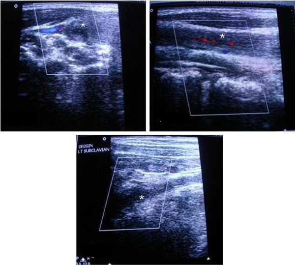
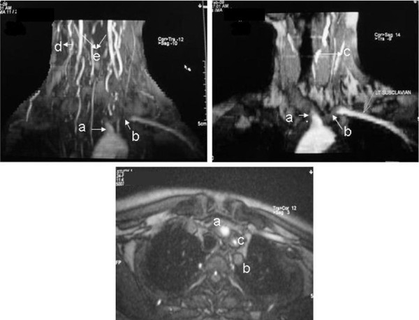
Case presentation: A 19-year-old woman of Asian origin was referred to our neurology out-patient department for the management of refractory seizures. She reported several episodes of a loss of consciousness with tonic posturing when she assumed an upright position, which was accompanied by constitutional symptoms. A clinical examination showed orthostatic hypotension and an investigation confirmed the diagnosis of Takayasu's disease with presentation as convulsive syncope.
Conclusion: Our case highlights the importance of a thorough clinical history and physical examination in order to distinguish events mimicking epileptic seizure. We also describe an unusual presentation of Takayasu's disease with convulsive syncope and systemic constitutional symptoms.
Figures
Similar articles
-
Refractory convulsive syncope in pregnancy: a rare presentation of Takayasu's arteritis - a case report and literature review.Afr Health Sci. 2021 Jun;21(2):852-857. doi: 10.4314/ahs.v21i2.46. Afr Health Sci. 2021. PMID: 34795744 Free PMC article. Review.
-
Takayasu's Arteritis presenting as a dissecting aortic aneurysm history: a case report.Cases J. 2008 Jul 21;1(1):52. doi: 10.1186/1757-1626-1-52. Cases J. 2008. PMID: 18644125 Free PMC article.
-
Clinical, angiographic profile and percutaneous endovascular management of Takayasu's arteritis - A single centre experience.Int J Cardiol. 2016 Oct 1;220:924-8. doi: 10.1016/j.ijcard.2016.06.194. Epub 2016 Jun 26. Int J Cardiol. 2016. PMID: 27420344
-
An unusual case of Takayasu's arteritis: Evaluation by CT angiography.Ann Indian Acad Neurol. 2011 Oct;14(4):304-6. doi: 10.4103/0972-2327.91960. Ann Indian Acad Neurol. 2011. PMID: 22346024 Free PMC article.
-
Takayasu's arteritis: a cell-mediated large-vessel vasculitis.Int J Clin Lab Res. 1999;29(1):8-13. doi: 10.1007/s005990050055. Int J Clin Lab Res. 1999. PMID: 10356657 Review.
Cited by
-
Takayasu Arteritis Mistaken for Epilepsy: A Case Presenting With Convulsive Syncope.J Med Cases. 2020 Feb;11(2):37-40. doi: 10.14740/jmc3424. Epub 2020 Feb 28. J Med Cases. 2020. PMID: 34434357 Free PMC article.
References
-
- Nagasawa T. Current status of large and small vessel vasculitis in Japan. Int J Cardiol. 1998;54:S98. - PubMed
LinkOut - more resources
Full Text Sources
