. 2010 Nov 2:5:30.
doi: 10.1186/1749-8104-5-30.

Pro-neurotrophins secreted from retinal ganglion cell axons are necessary for ephrinA-p75NTR-mediated axon guidance

Affiliations

Pro-neurotrophins secreted from retinal ganglion cell axons are necessary for ephrinA-p75NTR-mediated axon guidance

Katharine J M Marler et al. Neural Dev. .

Abstract

Background: Retinotectal map formation develops via topographically specific guidance and branching of retinal axons in their target area. This process is controlled, in part, by reverse signalling of ephrinAs expressed on retinal axons. As glycosylphosphatidylinositol-anchored molecules, ephrinAs require transmembrane co-receptors to exert this function, for which the two neurotrophin receptors, p75NTR and TrkB, were recently proposed.

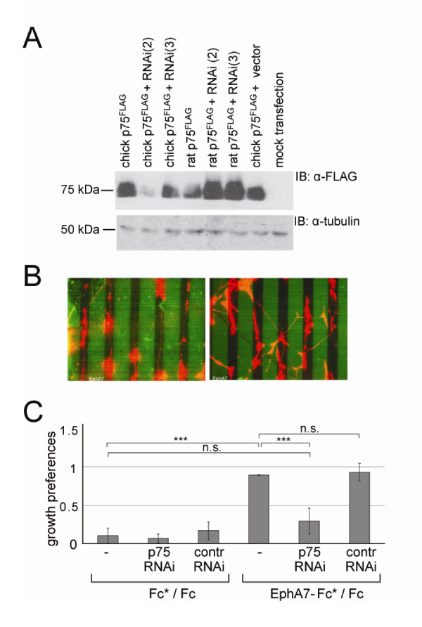
Results: We show here that the ligands for these receptors, the brain-derived neurotrophic factor precursor (proBDNF) and its processed form, BDNF, respectively, control the branching of retinal axons antagonistically, which they mediate by inducing the corresponding neurotrophin receptor-ephrinA complexes. Moreover, scavenging proneurotrophins, by adding antibodies specific for the pro-domain of proBNDF or a soluble extracellular domain of p75NTR, abolish repellent ephrinA reverse signalling in the stripe assay.

Conclusions: This indicates that retinal cells secrete proneurotrophins, inducing the ephrinA-p75NTR interaction and enabling repellent axon guidance. The antagonistic functions of proBDNF and BDNF raise the possibility that topographic branching is controlled by local control of processing of proneurotrophins.


Figures

Figure 1
Figure 1
The ephrinA5-p75NTR interaction is promoted in a ligand-dependent manner. CHO cells were transfected with cDNA expression vectors for p75NTR, FLAG and ephrinA5HA. A day later cells were serum starved and treated for 30 minutes with 100 ng/ml NGF as indicated. Subsequently, cells were lysed and immunoprecipitated using a αHA antibody. Western blot analyses of input and immunoprecipitate showed that co-immunoprecipitation of p75NTR and ephrinA5 is increased in the presence of ligand. A quantification of these experiments and comparable experiments using proneurotrophins are shown in Additional files 1 and 2. The asterisk marks the Ig heavy chain of the antibody used for immunoprecipitation. IB, immunoblot; IP, immunoprecipitation.
Figure 2
Figure 2
Guidance of retinal axons on an EphA7-Fc versus Fc substrate is abolished by knockdown of p75NTR. (A) Efficiency of selected RNAi experiments to knock down p75NTR protein. Chick or rat p75NTR, FLAG was expressed in CHO cells in parallel to different RNAi experiments targeting selected chick p75NTR sequences. RNAi(2) almost completely knocks down chick but not rat p75NTR, while RNAi(3) has no effect on either. α-Tubulin levels are shown for loading controls. (B) Single cells from embryonic day 6 nasal thirds of chick retina were electroporated with RNAi vectors for p75NTR. These RNAi vectors also contained a red fluorescent protein (RFP) cassette. Then cells were plated on a substrate of alternating lanes of EphA7-Fc and Fc stripes. The first generated stripe was labelled by adding FITC-Fc at low concentrations (indicated by EphA7-Fc*, or Fc* in Fc/Fc controls). In the pictures shown, the green stripes represent the EphA7-Fc stripe. Stripe width is 50 μm. Two days later, outgrowth preferences of retinal axons were analysed. The picture on the left shows the growth preference on EphA7-Fc*/Fc stripes after electroporation of control RNAi(3), and the picture on the right that after electroporation of RNAi(2). Knockdown of p75NTR leads to a strong abolishment of the striped outgrowth of retinal axons. For all stripe assays (and outgrowth/branching assays) the evaluation was done blind to the composition of stripes, constructs transfected and ligands added. (C) Quantification of growth preferences after p75NTR knockdown. The results of three independent experiments are shown. Error bars denote standard error of the mean and significance is indicated as ***P < 0.001; n.s., not significant. Statistical analysis was done in GraphPad using one way ANOVA and Tukey post hoc test (GraphPad: LaJolla, CA, USA).
Figure 3
Figure 3
Expression of proBDNF in chick RGC axons. Retinal single cell cultures derived from E6 chick retina were stained after 2 days in vitro using a monoclonal antibody against the pro-domain of proBDNF [14] according to protocols given in Yang et al. [14]. (A-C) An RGC growth cone stained with control antibody (B), and phalloidin (C) to show the location of actin. (A) The composite of (B) and (C). In all composites phalloidin is shown in green and proBDNF/control antibody in red. (D-F) An RGC axon stained with control antibody (E), and phalloidin (F). (D) The composite of (E) and (F). (G-I) An RGC axon stained with a proBDNF antibody (H) [14], and phalloidin (I). (G) The composite of (H) and (I). (J-L). An RGC growth cone stained with proBDNF antibody (K), and phalloidin (L). (J) The composite of (K) and (L).
Figure 4
Figure 4
Repellent axon guidance is disrupted in the presence of an anti-proBDNF antibody or a soluble extracellular domain of p75NTR. (A) Quantification of axon growth preferences in the presence of an anti-proBDNF antibody [14] or a control antibody. Stripe assay experiments were performed in the presence of a proBDNF antibody (1:200) [14] or a control antibody (mouse monoclonal antibody for placental alkaline phosphatase; 1:200). The quantification of axon growth preferences shows an abolishment of repellent guidance in the presence of the proBDNF antibody (see also Additional file 4). Error bars represent the standard error of the mean. Statistics were performed using Kruskal-Wallis test and Dunn's multiple comparison test with ***P < 0.001. (B) Immunoprecipitation and western blot analysis of CHO cell supernatants transfected with p75NTR, sol, FLAG analysing two different clones (c7 and c9). The arrowhead points to the band corresponding to p75NTR, sol, FLAG; the asterisk indicates the Ig light chain from the αFLAG antibody. The first lane shows the analysis of mock-transfected cells. IB, immunoblot; IP, immunoprecipitation. (C-F) Single cells from E6 retina were electroporated with an eGFP expression plasmid and plated on alternating lanes of EphA7-Fc/Fc, or Fc/Fc (see (A)). Two days later, the cultures were fixed and analysed for their growth preferences. (C, E) Fc versus Fc stripes. (D, F) EphA7-Fc versus Fc stripes. (C, D) Addition of media from mock transfected CHO cells. (E, F) Addition of media from CHO cells transfected with an expression plasmid for the extracellular soluble domain of p75NTR (p75NTR, sol). (G) Quantification of axon growth preferences in the presence of p75NTR, sol or control medium. Two different p75NTR, sol, FLAG clones (c7 and c9) were analysed and led to a similar reduction in the growth preference of RGC axons. Statistics were performed in GraphPad using Kruskal-Wallis with post hoc Dunn's multiple comparison test.
Figure 5
Figure 5
Proneurotrophins abolish retinal axon branching via p75NTR. The outgrowth/branching assay was performed as described [6]. Cells from E8 nasal retina were electroporated with eGFP and plated on a merosin/laminin substrate. (A) Cultures were treated at 1 day in vitro with 5 ng/ml BDNF, or 5 ng/ml proneurotrophins in the presence of 5 ng/ml BDNF as indicated, and fixed and analysed for branch number per axon after 3 days in vitro. The basal level of branching was not affected by treatment of retinal cultures with proneurotrophins alone. However, both proBDNF and proNGF led to a downregulation of the BDNF-induced branching to basal levels. (B) The length of outgrowth of retinal axons is not affected by treatment with neurotrophins and/or proneurotrophins. Axon length is given in arbitrary units. (C) To show that the proBDNF effect is mediated via p75NTR, retinal cultures were electroporated either with an RNAi vector resulting in the knockdown of p75NTR, with an RNAi vector not affecting p75NTR protein levels or with empty vector. After plating, the cultures were treated with pro/neurotrophins as described in (A). p75NTR knockdown obliterates the branch-suppressing effect of proBDNF. Three independent experiments were performed. The statistical analysis was done using Kruskal-Wallis test and Dunn's multiple comparison test post hoc. ***P < 0.001; n.s., not significant, the error bars represent S.E, M.

Similar articles

Cited by

References

    1. Clandinin TR, Feldheim DA. Making a visual map: mechanisms and molecules. Curr Opin Neurobiol. 2009;19:174–180. doi: 10.1016/j.conb.2009.04.011. - DOI - PMC - PubMed
    1. Huberman AD, Feller MB, Chapman B. Mechanisms underlying development of visual maps and receptive fields. Annu Rev Neurosci. 2008;31:479–509. doi: 10.1146/annurev.neuro.31.060407.125533. - DOI - PMC - PubMed
    1. McLaughlin T, O'Leary DD. Molecular gradients and development of retinotopic maps. Annu Rev Neurosci. 2005;28:327–355. doi: 10.1146/annurev.neuro.28.061604.135714. - DOI - PubMed
    1. Rashid T, Upton AL, Blentic A, Ciossek T, Knoll B, Thompson ID, Drescher U. Opposing gradients of ephrin-As and EphA7 in the superior colliculus are essential for topographic mapping in the mammalian visual system. Neuron. 2005;47:57–69. doi: 10.1016/j.neuron.2005.05.030. - DOI - PubMed
    1. Lim YS, McLaughlin T, Sung TC, Santiago A, Lee KF, O'Leary DD. p75(NTR) mediates ephrin-A reverse signaling required for axon repulsion and mapping. Neuron. 2008;59:746–758. doi: 10.1016/j.neuron.2008.07.032. - DOI - PMC - PubMed

Publication types

MeSH terms