. 2010 Nov 2:7:91.
doi: 10.1186/1742-4690-7-91.

BLV-CoCoMo-qPCR: Quantitation of bovine leukemia virus proviral load using the CoCoMo algorithm

Affiliations

BLV-CoCoMo-qPCR: Quantitation of bovine leukemia virus proviral load using the CoCoMo algorithm

Mayuko Jimba et al. Retrovirology. .

Abstract

Background: Bovine leukemia virus (BLV) is closely related to human T-cell leukemia virus (HTLV) and is the etiological agent of enzootic bovine leukosis, a disease characterized by a highly extended course that often involves persistent lymphocytosis and culminates in B-cell lymphomas. BLV provirus remains integrated in cellular genomes, even in the absence of detectable BLV antibodies. Therefore, to understand the mechanism of BLV-induced leukemogenesis and carry out the selection of BLV-infected animals, a detailed evaluation of changes in proviral load throughout the course of disease in BLV-infected cattle is required. The aim of this study was to develop a new quantitative real-time polymerase chain reaction (PCR) method using Coordination of Common Motifs (CoCoMo) primers to measure the proviral load of known and novel BLV variants in clinical animals.

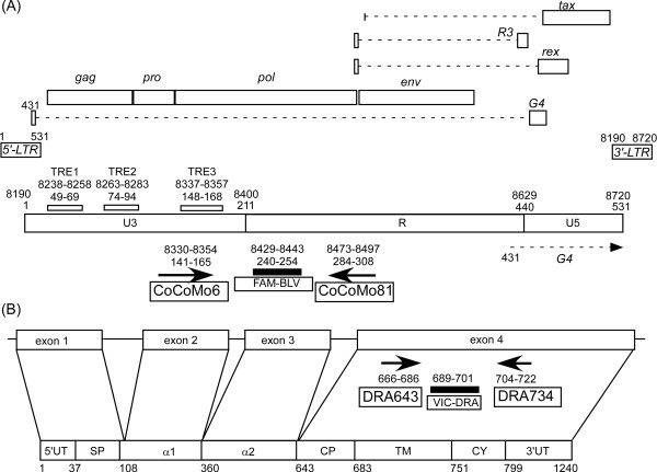
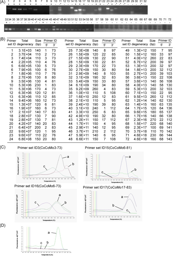
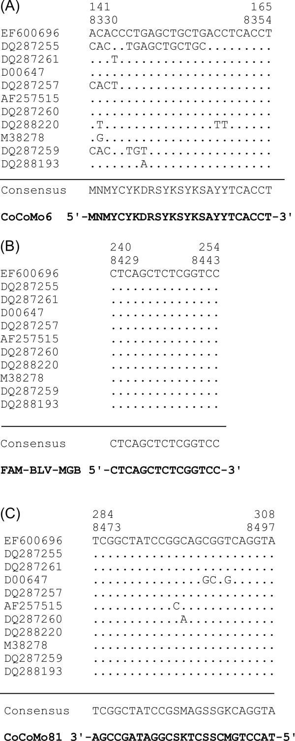
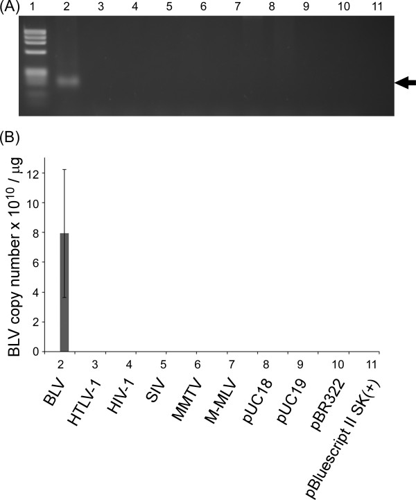
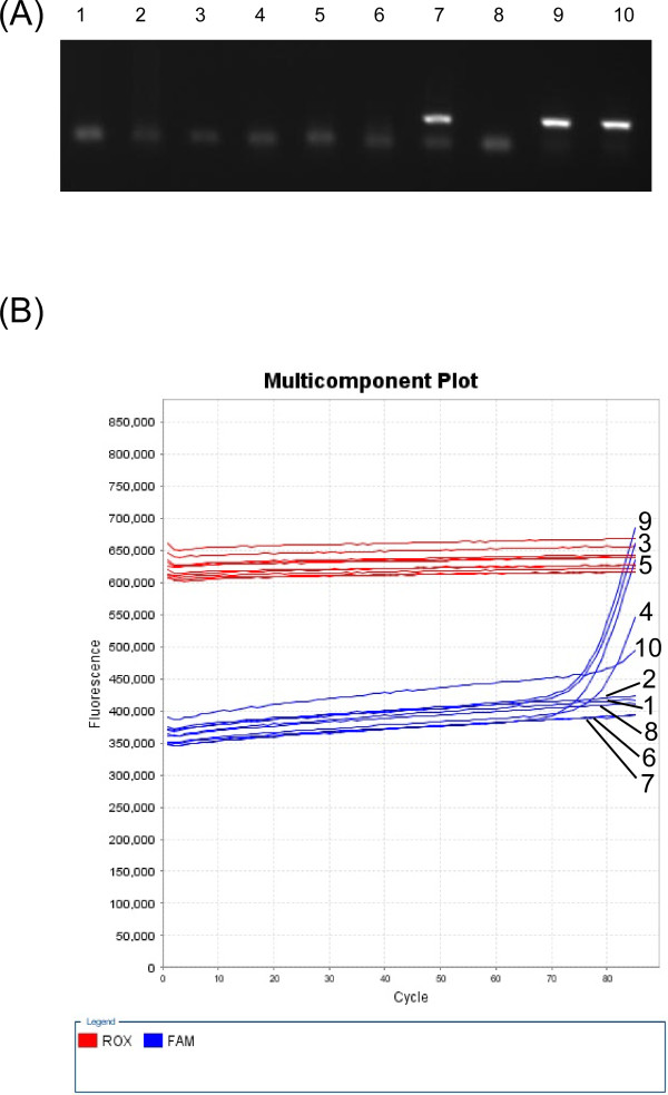
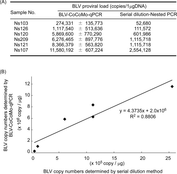
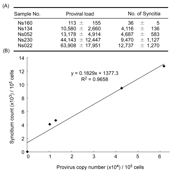
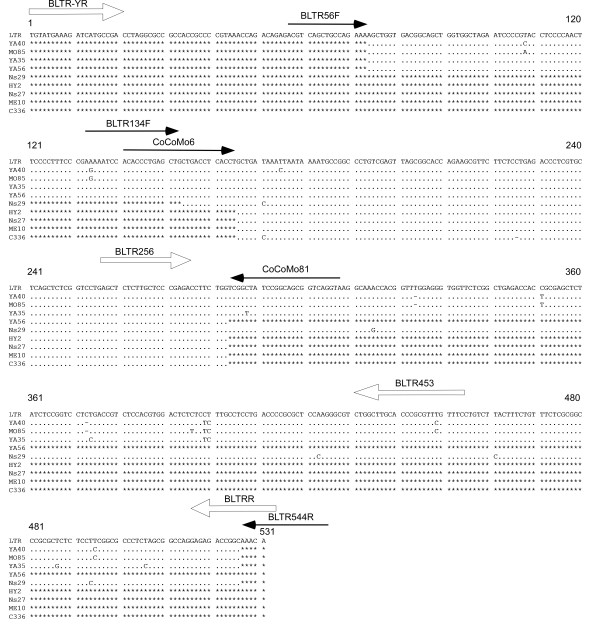
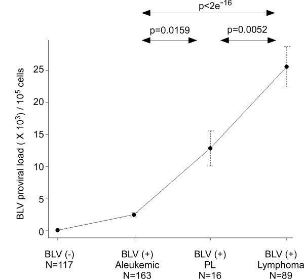
Results: Degenerate primers were designed from 52 individual BLV long terminal repeat (LTR) sequences identified from 356 BLV sequences in GenBank using the CoCoMo algorithm, which has been developed specifically for the detection of multiple virus species. Among 72 primer sets from 49 candidate primers, the most specific primer set was selected for detection of BLV LTR by melting curve analysis after real-time PCR amplification. An internal BLV TaqMan probe was used to enhance the specificity and sensitivity of the assay, and a parallel amplification of a single-copy host gene (the bovine leukocyte antigen DRA gene) was used to normalize genomic DNA. The assay is highly specific, sensitive, quantitative and reproducible, and was able to detect BLV in a number of samples that were negative using the previously developed nested PCR assay. The assay was also highly effective in detecting BLV in cattle from a range of international locations. Finally, this assay enabled us to demonstrate that proviral load correlates not only with BLV infection capacity as assessed by syncytium formation, but also with BLV disease progression.

Conclusions: Using our newly developed BLV-CoCoMo-qPCR assay, we were able to detect a wide range of mutated BLV viruses. CoCoMo algorithm may be a useful tool to design degenerate primers for quantification of proviral load for other retroviruses including HTLV and human immunodeficiency virus type 1.


Figures

Figure 1
Figure 1
The position, length and orientation of primers and probes used in the bovine leukemia virus (BLV)-CoCoMo-qPCR method. Labeled arrows indicate the orientation and length of each primer. The black filled box indicates the probe annealing position. (A) The proviral structure of BLV in the BLV cell line FLK-BLV subclone pBLV913, complete genome [DDBJ: EF600696]. It contains two LTR regions at nucleotide positions 1-531 and 8190-8720. Lowercase labels indicate these LTR regions. The upper number shows the position of the 5' LTR and the lower number shows the position of the 3'LTR. Both LTRs include the U3, R and U5 regions. A triplicate 21-bp motif known as the Tax-responsive element (TRE) is present in the U3 region of the 5' LTR. The target region for amplification was in the U3 and R region, and the TaqMan probe for detecting the PCR product was from the R region. (B) The schematic outline of the bovine major histocompatibility complex (BoLA)-DRA gene (upper) and its cDNA clone MR1 [DDBJ: D37956] (lower). Exons are shown as open boxes. The numbers indicate the numbering of the nucleotide sequence of MR1. 5'UT, 5'-untranslated region; SP, signal sequence; α1, first domain; α 2, second domain; CP, connecting peptide; TM, transmembrane domain; CY, cytoplasmic domain; 3'UT, 3-untranslated region. The target regions for amplification and for binding of the TaqMan probe to detect the PCR product are in exon 4.
Figure 2
Figure 2
Selection of the primer set for amplification of the BLV LTR region. (A) Touch-down PCR was performed using 72 primer sets with 49 primers designed by the CoCoMo program as shown in Table 1. PCR products were detected by electrophoresis on a 3% agarose gel. Lanes 1-72, 1-72 primer set ID; +, results positive for PCR product; -, negative results for same. *, designates PCR products that were detected but for which the amplicon sizes differed from the predicted size. (B) Summary of results shown in (A). Primer set IDs are arranged according to the degeneracy of the primer set and size of the PCR products. (C) The 4 representative melting curves with 16 primer sets of: BLV-infected BLSC-KU-17 cells (a), BLV-free normal cattle cells (b), and reagent-only as negative control (c). The specificity of the 16 selected primer sets was checked by melting curve analysis. Each PCR amplification was followed by gradual product melting at up to 95°C. (D) The optimization of PCR amplification with primer set ID 15 (CoCoMo 6 and 81). The melting curve of PCR products from BLV-infected BLSC-KU-17 cells (a), the BLV-free normal cattle Ns118 (b), and reagent-only as negative control (c).
Figure 3
Figure 3
Sequence alignment of annealing positions of the CoCoMo 6 primer (A), FAM-BLV-MGB probe (B) and CoCoMo 81 primer (C) in the 52 BLV LTR sequences. The sequence alignment used 52 sequences from GenBank that were integrated into a total of 11 sequences, including 8 individual sequences for the CoCoMo 6 primer and 4 individual sequences for the CoCoMo81 primer. Accession numbers for the representative sequences are indicated in the left column. Numbers indicate the numbering of the nucleotide sequence of the FLK-BLV subclone pBLV913 [DDBJ: EF600696]. The upper number shows the position of the 5' LTR and the lower number shows the position of the 3' LTR.
Figure 4
Figure 4
Evaluation of the specificity of the BLV-CoCoMo-qPCR primers. (A) Real-time PCR using the CoCoMo 6 and CoCoMo 81 primers from the BLV-CoCoMo-qPCR was performed using 0.3 ng of the following infectious molecular clones: BLV (pBLV-IF, lane 2); HTLV-1 (pK30, lane 3); HIV-1 (pNL4-3, lane 4); SIV (pSIVmac239/WT, lane 5); MMTV (hybrid MMTV, lane 6); M-MLV (pL-4, lane 7); and the plasmids pUC18 (lane 8), pUC19 (lane 9), pBR322 (lane 10), and pBluescript SK(+) (lane 11). PCR products were subjected to 3% agarose gel electrophoresis. Lane 1, DNA marker Φ × 174-Hae III digest. A PCR product 168 bp in length is indicated by an arrow. (B) The number of BLV provirus copies in 1 μg of DNA from each DNA sample is indicated by lowercase. Values represent the mean ± standard deviation (SD) of the results of three independent experiments.
Figure 5
Figure 5
Comparison of the sensitivity of BLV-CoCoMo-qPCR and nested PCR. Ten samples containing 0.7 copies of pBLV-LTR/SK were amplified by nested PCR (A) and real-time PCR with the CoCoMo 6 and CoCoMo 81 primer set (B). The 168-bp band was used to detect BLV LTR amplicons (A). Carboxy-X-rhodamine (ROX) intensities were used for corrections of tube differences, and carboxyfluorescein (FAM) intensities were used to detect BLV LTR amplicons (B).
Figure 6
Figure 6
Correlation between proviral load calculated by BLV-CoCoMo-qPCR and serial dilution nested PCR. (A) BLV proviral copy numbers for 1 μg of genomic DNA from 6 BLV-infected cattle were determined by BLV-CoCoMo-qPCR and serial dilution-nested PCR. For serial dilution-nested PCR, the 6 genomic DNAs were analyzed by serial tenfold dilution and subjected to nested PCR for detection of the BLV LTR gene. Nested PCR reactions were repeated 10 times and proviral load was calculated according to a Poisson distribution model as shown in Methods. Values represent the mean ± standard deviation (SD) of results from four independent experiments. (B) Scatter chart is indicated the correlation between BLV copy numbers which were determined by BLV-CoCoMo-qPCR and by serial dilution-nested PCR.
Figure 7
Figure 7
Correlation between proviral load calculated by BLV-CoCoMo-qPCR and syncytium formation. (A) Using BLV-CoCoMo-qPCR, the proviral loads from five BLV-infected cattle were calculated and shown as provirus copy number per 1 × 10⁵ cells. A syncytium formation assay using CC81 indicator cells was used to count the number of syncytia per 1 × 10⁵ peripheral blood mononuclear cells (PBMCs) from five BLV-infected cattle. Values represent the mean ± standard deviation (SD) of results from three samples. (B) Scatter chart is indicated the correlation between BLV copy numbers which were determined by BLV-CoCoMo-qPCR and the number of syncytia.
Figure 8
Figure 8
Alignment of BLV LTR nucleotide sequences in samples that were positive by BLV-CoCoMo-qPCR but negative by nested PCR. BLV LTR sequences from 9 BLV-infected cattle were amplified by PCR using three primer pairs, BLTR56F and CoCoMo81, BLTR134F and BLTR544R, and CoCoMo6 and CoCoMo81, followed by sequencing of the PCR products. Closed arrows indicate the position, orientation and length of these primers. The LTR sequences at nucleotide positions 74-283 and 154-526 from YA40, MO85 and YA35 were amplified using two primer pairs BLTR56F and CoCoMo81, and BLTR134F and BLTR544R, respectively. The LTR sequence at nucleotide positions 74-283 from YA56 was amplified by primer pair BLTR56F and CoCoMo81. The LTR sequences at nucleotide positions 166-283 from HY2, Ns27, ME10 and C336 were amplified by the primer pair CoCoMo6 and CoCoMo81. The LTR sequence at nucleotide positions 154-526 from Ns29 was amplified by primer pair BLTR134F and BLTR544R. Open arrows indicate the position, orientation and length of first primer pair, BLTR-YR and BLTRR, and the second primer pair, BLTR256 and BLTR453, for nested PCR. The numbers at the top of the sequences indicate the terminal bases according to the nucleotide sequence of the FLK-BLV subclone pBLV913 [DDBJ: EF600696]. Conserved sequences are indicated by a dot (.), deletions are indicated by a hyphen (-). A lack of sequence information is indicated by the symbol (*).
Figure 9
Figure 9
Increased proviral load correlates with disease progression in BLV-induced enzootic bovine leukosis (EBL). The proviral load was calculated for 385 cattle by BLV-CoCoMo-qPCR. The cattle were classified into four disease stages according to diagnosis based on previously established criteria [41], the genomic integration of BLV, and the detection of antibodies to BLV: 117 BLV-negative cattle (BLV-); 163 BLV-infected cattle that were clinically and hematologically normal (aleukemic); 16 clinically normal, BLV-infected cattle with persistent lymphocytosis (PL); and 89 BLV-infected cattle with lymphoma. The circles/dots indicate the average proviral load detected in each stage, and the bar indicates the standard error. P-values were calculated by pairwise t-test using R version 2.10.1 (The R Foundation for Statistical Computing).

Similar articles

Cited by

References

    1. Lloyd-Smith JO, George D, Pepin KM, Pitzer VE, Pulliam JR, Dobson AP, Hudson PJ, Grenfell BT. Epidemic dynamics at the human-animal interface. Science. 2009;326:1362–1367. doi: 10.1126/science.1177345. - DOI - PMC - PubMed
    1. Tapper ML. Emerging viral diseases and infectious disease risks. Haemophilia. 2006;12(Suppl 1):3–7. doi: 10.1111/j.1365-2516.2006.01194.x. discussion 26-28. - DOI - PMC - PubMed
    1. Endoh D, Mizutani T, Kirisawa R, Maki Y, Saito H, Kon Y, Morikawa S, Hayashi M. Species-independent detection of RNA virus by representational difference analysis using non-ribosomal hexanucleotides for reverse transcription. Nucleic Acids Res. 2005;33:e65. doi: 10.1093/nar/gni064. - DOI - PMC - PubMed
    1. Rose TM, Schultz ER, Henikoff JG, Pietrokovski S, McCallum CM, Henikoff S. Consensus-degenerate hybrid oligonucleotide primers for amplification of distantly related sequences. Nucleic Acids Res. 1998;26:1628–1635. doi: 10.1093/nar/26.7.1628. - DOI - PMC - PubMed
    1. Bartl S. Amplification using degenerate primers with multiple inosines to isolate genes with minimal sequence similarity. Methods Mol Biol. 1997;67:451–457. - PubMed

Publication types

MeSH terms

LinkOut - more resources