. 2010 Nov 2:10:274.
doi: 10.1186/1471-2180-10-274.

Genetic analysis of the capsule polysaccharide (K antigen) and exopolysaccharide genes in pandemic Vibrio parahaemolyticus O3:K6

Affiliations

Genetic analysis of the capsule polysaccharide (K antigen) and exopolysaccharide genes in pandemic Vibrio parahaemolyticus O3:K6

Yuansha Chen et al. BMC Microbiol. .

Abstract

Background: Pandemic Vibrio parahaemolyticus has undergone rapid changes in both K- and O-antigens, making detection of outbreaks more difficult. In order to understand these rapid changes, the genetic regions encoding these antigens must be examined. In Vibrio cholerae and Vibrio vulnificus, both O-antigen and capsular polysaccharides are encoded in a single region on the large chromosome; a similar arrangement in pandemic V. parahaemolyticus would help explain the rapid serotype changes. However, previous reports on "capsule" genes are controversial. Therefore, we set out to clarify and characterize these regions in pandemic V. parahaemolyticus O3:K6 by gene deletion using a chitin based transformation strategy.

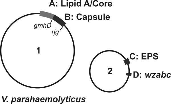
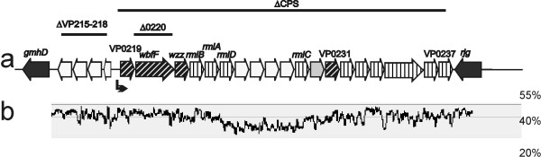
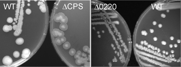
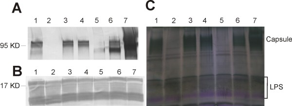
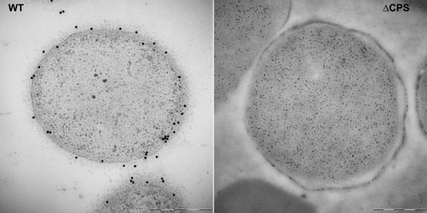
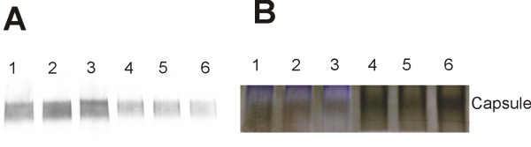
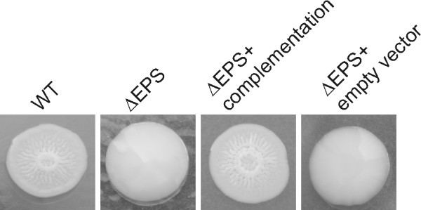
Results: We generated different deletion mutants of putative polysaccharide genes and examined the mutants by immuno-blots with O and K specific antisera. Our results showed that O- and K-antigen genes are separated in V. parahaemolyticus O3:K6; the region encoding both O-antigen and capsule biosynthesis in other vibrios, i.e. genes between gmhD and rjg, determines the K6-antigen but not the O3-antigen in V. parahaemolyticus. The previously identified "capsule genes" on the smaller chromosome were related to exopolysaccharide synthesis, not K-antigen.

Conclusion: Understanding of the genetic basis of O- and K-antigens is critical to understanding the rapid changes in these polysaccharides seen in pandemic V. parahaemolyticus. This report confirms the genetic location of K-antigen synthesis in V. parahaemolyticus O3:K6 allowing us to focus future studies of the evolution of serotypes to this region.


Figures

Figure 1
Figure 1
Gene clusters related to polysaccharide in Vibrio parahaemolyticus O3:K6. Two circles to represent two chromosomes. Function of each region is indicated. A (VP0190-0214), putative lipid A/core region; B (VP0215-0237), K-antigen/capsule region (CPS); C (VPA1403-1412), exopolysaccharide region (EPS); D (VPA1602-1604), putative polysaccharide exportation genes wza, b, c.
Figure 2
Figure 2
Capsule (K-antigen) genes in V. parahaemolyticus O3:K6. a) Bars with mutant names above indicate regions deleted in each mutant. Bent arrow indicates promoter. Design patterns of open reading frames indicate different classes of genes: vertical lines, pathway genes; diagonal lines, processing and transportation genes; grey box, glycosyltransferase; white box, functions not clear. b) GC percentage of the sequence in 120 bp windows, aligned to the genes in a.
Figure 3
Figure 3
V. parahaemolyticus mutants ∆CPS and ∆0220 display translucent phenotype. Wild type (WT), ∆CPS and ∆0220 have grown on LB agar at 37°C for 24 hours.
Figure 4
Figure 4
Immunoblots and stains-all/silver-stain of V. parahaemolyticus. Whole cells lysate treated with DNase, RNase and pronase was separated on polyacrylamide gel, transferred to PVDF membrane and probed with K6 specific antiserum (A), or O3 specific antiserum (B). Total polysaccharides were visualized by stains-all/silver-stain on polyacrylamide gel (C). lane 1, wild type VP53; lane 2, ∆CPS mutant; lane 3, ∆EPS mutant; lane 4, ∆wzabc mutant; lane 5, ∆0220 mutant; lane 6, ∆0220 mutant with trans-complementation; lane 7, ∆VP215-218 mutant.
Figure 5
Figure 5
Immuno-gold labeling TEM of V. parahaemolyticus with K6 antiserum. Thin sections samples were labeled with K6 antiserum, followed by gold attached secondary antibodies. Left, Wild type VP53 (WT), right, ∆CPS mutant. Bar equals to 500 nm.
Figure 6
Figure 6
Immuno blot and stains-all/silver-stain of cell fractions. Outer membrane (OM) and cytoplasmic (CP) fractions were separated on polyacrylamide gel, then were either transferred to PVDF membrane and probed with K6 specific antiserum (A), or stained with stains-all/silver stain (B). Lane1, wild type CP; lane 2, ∆wzabc CP; lane 3, ∆EPS CP; lane 4, wild type OM; lane 5, ∆wzabc OM; lane 6, ∆EPS OM.
Figure 7
Figure 7
Colony morphology of V. parahaemolyticus. Wild type (WT) V. parahaemolyticus displayed rugose phenotype when incubated in APW#3 media followed by 48-72 hours incubation on LB agar. Mutant ∆EPS only displayed smooth phenotype under the same conditions. Complementation of ∆EPS by the EPS genes restored the rugose phenotype while the ∆EPS mutant with empty vector remained smooth.

References

    1. Fujino L, Okuno Y, Nakada D, Aoyama A, Fukai K, Mukai T, Uebo T. On the bacteriological examination of shirasu food poisoning. Med J Osaka Univ. 1953;4:299–304.
    1. Nair GB, Ramamurthy T, Bhattacharya SK, Dutta B, Takeda Y, Sack DA. Global dissemination of Vibrio parahaemolyticus serotype O3:K6 and its serovariants. Clin Microbiol Rev. 2007;20(1):39–48. doi: 10.1128/CMR.00025-06. - DOI - PMC - PubMed
    1. Nair GB, Hormazabal JC. The Vibrio parahaemolyticus pandemic. Rev Chilena Infectol. 2005;22(2):125–130. - PubMed
    1. Chowdhury NR, Chakraborty S, Ramamurthy T, Nishibuchi M, Yamasaki S, Takeda Y, Nair GB. Molecular evidence of clonal Vibrio parahaemolyticus pandemic strains. Emerg Infect Dis. 2000;6(6):631–636. doi: 10.3201/eid0606.000612. - DOI - PMC - PubMed
    1. Chowdhury NR, Stine OC, Morris JG, Nair GB. Assessment of evolution of pandemic Vibrio parahaemolyticus by multilocus sequence typing. J Clin Microbiol. 2004;42(3):1280–1282. doi: 10.1128/JCM.42.3.1280-1282.2004. - DOI - PMC - PubMed

MeSH terms

LinkOut - more resources