

Syndecan-1 knock-down in decidualized human endometrial stromal cells leads to significant changes in cytokine and angiogenic factor expression patterns
- PMID: 21044331
- PMCID: PMC2988802
- DOI: 10.1186/1477-7827-8-133
Syndecan-1 knock-down in decidualized human endometrial stromal cells leads to significant changes in cytokine and angiogenic factor expression patterns
Abstract
Background: Successful embryonic implantation depends on a synchronized embryo-maternal dialogue. Chemokines, such as chemokine ligand 1 (CXCL1), play essential roles in the maternal reproductive tract leading to morphological changes during decidualization, mediating maternal acceptance towards the semi-allograft embryo and induction of angiogenesis. Chemokine binding to their classical G-protein coupled receptors is essentially supported by the syndecan (Sdc) family of heparan sulfate proteoglycans. The aim of this study was to identify the involvement of Sdc-1 at the embryo-maternal interface regarding changes of the chemokine and angiogenic profile of the decidua during the process of decidualization and implantation in human endometrium.
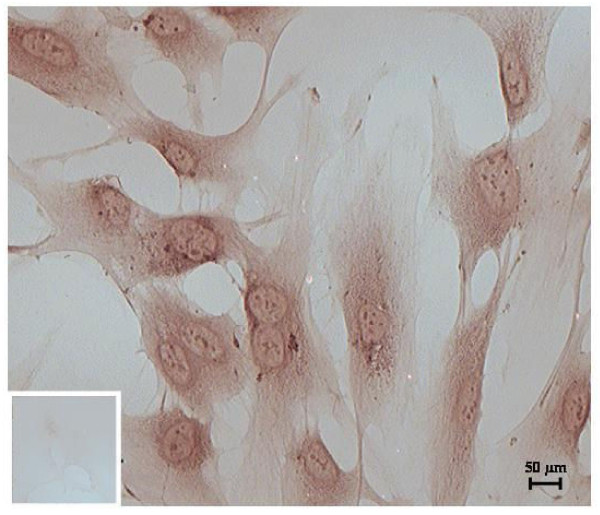
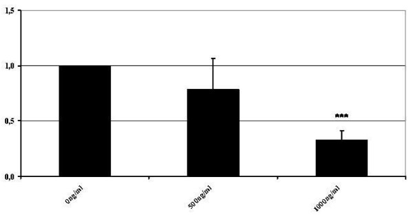
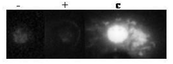
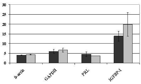
Methods: A stable Sdc-1 knock-down was generated in the immortalized human endometrial stromal cell line St-T1 and was named KdS1. The ability of KdS1 to decidualize was proven by Insulin-like growth factor binding 1 (IGFBP1) and prolactin (PRL) confirmation on mRNA level before further experiments were carried out. Dot blot protein analyses of decidualized knock-down cells vs non-transfected controls were performed. In order to imitate embryonic implantation, decidualized KdS1 were then incubated with IL-1beta, an embryo secretion product, vs controls. Statistical analyses were performed applying the Student's t-test with p < 0.05, p < 0.02 and p < 0.01 and one way post-hoc ANOVA test with p < 0.05 as cut-offs for statistical significance.
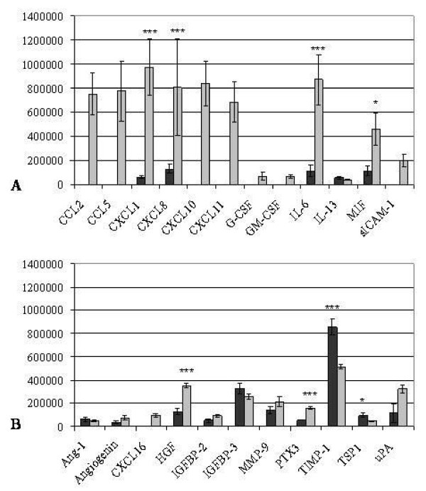
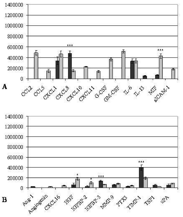
Results: The induction of the Sdc-1 knock-down revealed significant changes in cytokine and angiogenic factor expression profiles of dKdS1 vs decidualized controls. Incubation with embryonic IL-1beta altered the expression patterns of KdS1 chemokines and angiogenic factors towards inflammatory-associated molecules and factors involved in matrix regulation.
Conclusions: Sdc-1 knock-down in human endometrial stroma cells led to fulminant changes regarding cytokine and angiogenic factor expression profiles upon decidualization and imitation of embryonic contact. Sdc-1 appears to play an important role as a co-receptor and storage factor for many cytokines and angiogenic factors during decidualization and implantation period, supporting proper implantation and angiogenesis by regulation of chemokine and angiogenic factor secretion in favour of the implanting embryo.
Figures
References
-
- McEwan M, Lins RJ, Munro SK, Vincent ZL, Ponnampalam AP, Mitchell MD. Cytokine regulation during the formation of the fetal-maternal interface: Focus on cell-cell adhesion and remodelling of the extra-cellular matrix. Cytokine Growth factor Rev. 2009;20:241–249. doi: 10.1016/j.cytogfr.2009.05.004. - DOI - PubMed
Publication types
MeSH terms
Substances
LinkOut - more resources
Full Text Sources
Miscellaneous
