. 2010 Nov 2:6:74.
doi: 10.1186/1744-8069-6-74.

Peripheral nerve injury and TRPV1-expressing primary afferent C-fibers cause opening of the blood-brain barrier

Affiliations

Peripheral nerve injury and TRPV1-expressing primary afferent C-fibers cause opening of the blood-brain barrier

Simon Beggs et al. Mol Pain. .

Abstract

Background: The blood-brain barrier (BBB) plays the crucial role of limiting exposure of the central nervous system (CNS) to damaging molecules and cells. Dysfunction of the BBB is critical in a broad range of CNS disorders including neurodegeneration, inflammatory or traumatic injury to the CNS, and stroke. In peripheral tissues, the vascular-tissue permeability is normally greater than BBB permeability, but vascular leakage can be induced by efferent discharge activity in primary sensory neurons leading to plasma extravasation into the extravascular space. Whether discharge activity of sensory afferents entering the CNS may open the BBB or blood-spinal cord barrier (BSCB) remains an open question.

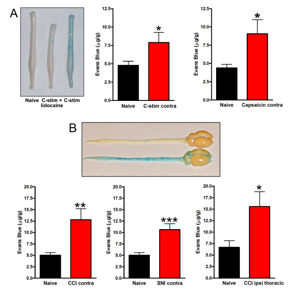
Results: Here we show that peripheral nerve injury (PNI) produced by either sciatic nerve constriction or transecting two of its main branches causes an increase in BSCB permeability, as assessed by using Evans Blue dye or horseradish peroxidase. The increase in BSCB permeability was not observed 6 hours after the PNI but was apparent 24 hours after the injury. The increase in BSCB permeability was transient, peaking about 24-48 hrs after PNI with BSCB integrity returning to normal levels by 7 days. The increase in BSCB permeability was prevented by administering the local anaesthetic lidocaine at the site of the nerve injury. BSCB permeability was also increased 24 hours after electrical stimulation of the sciatic nerve at intensity sufficient to activate C-fibers, but not when A-fibers only were activated. Likewise, BSCB permeability increased following application of capsaicin to the nerve. The increase in permeability caused by C-fiber stimulation or by PNI was not anatomically limited to the site of central termination of primary afferents from the sciatic nerve in the lumbar cord, but rather extended throughout the spinal cord and into the brain.

Conclusions: We have discovered that injury to a peripheral nerve and electrical stimulation of C-fibers each cause an increase in the permeability of the BSCB and the BBB. The increase in permeability is delayed in onset, peaks at about 24 hours and is dependent upon action potential propagation. As the increase is mimicked by applying capsaicin to the nerve, the most parsimonious explanation for our findings is that the increase in permeability is mediated by activation of TRPV1-expressing primary sensory neurons. Our findings may be relevant to the development of pain and neuroplastic changes in the CNS following nerve injury. In addition, our findings may provide the basis for developing methods to purposefully open the BBB when needed to increase brain penetration of therapeutic agents that might normally be excluded by an intact BBB.


Figures

Figure 1
Figure 1
Peripheral nerve injury (PNI) increases BSCB permeability. A: Increased permeability to Evans Blue of spinal cord from naïve rats, sham operated rats or rats with chronic constriction injury (CCI, left) or spared nerve injury (SNI, right). Inset: Standard curve of Evans blue and the representative reading of naïve rats or CCI rats. B: Increased permeability to horseradish peroxidase (HRP) following PNI. Left, representative spinal cord sections of HRP extravasation 24 hours after SNI, CCI or naïve rats. Right, histogram showing the integrated pixel density of HRP signaling. Data are presented as mean ± SEM; *p < 0.05 compared to naïve; n = 4-6 per group. All measurements are from lumbar spinal cord ipsilateral to PNI stimulation.
Figure 2
Figure 2
The effect of local lidocaine block and sequential nerve injury on CCI-induced increase in BSCB permeability. A: 2% of Lidocaine was applied to the sciatic nerve immediately following CCI, and Evans Blue extravasation in the ipsilateral lumbar spinal cord was measured 24 hours post CCI. B: A sequential CCI was performed on the right sciatic nerve after the initial CCI was performed in the left sciatic nerve 6 days previously. Evans blue extravasation was measured 7 days after initial CCI. All readings are from the left lumbar spinal cords (ipsilateral to the initial injury). Data are presented as mean ± SEM; *p < 0.05 **p < 0.01 compared to naïve; n = 5 per group.
Figure 3
Figure 3
C-fiber intensity stimulation increases BSCB permeability. A: Sciatic nerve stimulation with C-fiber intensity, but not A-fiber intensity increases permeability to Evans Blue in rat spinal cord 24 hours post stimulation. Data are presented as mean ± SEM; **p < 0.01 compared to naïve, n = 4-8 per group. B: Representative spinal cord sections of HRP extravasation from naïve rats or rats 24 hours post C-fiber intensity stimulation of the sciatic nerve. C: C-fiber intensity stimulation-induced permeability is blocked by pre-treatment, but not post-treatment, with lidocaine. 2% lidocaine was applied to the sciatic nerve either 30 minutes before C-fiber stimulation (pre-block) or immediately after C-fiber stimulation (post-block). The duration of lidocaine treatment was 30 minutes in both cases. Data are presented as mean ± SEM; **p < 0.01 compared to C-fiber stimulation, n = 4-8 per group. All the readings are from lumbar spinal cord ipsilateral to nerve stimulation.
Figure 4
Figure 4
Local application of capsaicin increases BSCB permeability. A: 1% Capsaicin or vehicle was applied onto the sciatic nerve and Evans Blue extravasation was measured 24 hours later. All readings are from lumbar spinal cord ipsilateral to capsaicin or vehicle. Data are presented as mean ± SEM; **p < 0.01 compared to naïve, n = 5-9 per group. B: 1% Capsaicin was applied to the sciatic nerve and Evans Blue was injected immediately after capsaicin. Ipsilateral and contralateral paw images were captured 45 minutes after capsaicin treatment.
Figure 5
Figure 5
Increased BSCB permeability extends throughout the spinal cord. A: left panel, representative spinal cord from naïve rats, rats 24 hours post C-fiber stimulation or rats with lidocaine block before C-fiber stimulation. Middle and right panels, C-fiber stimulation (middle) or local application of capsaicin onto sciatic nerve (right) increases contralateral spinal cord Evans Blue permeability 24 hours post treatment. B: Increased permeability to Evans Blue in the lumbar spinal cord contralateral to CCI (left panel) or SNI (middle) and in thoracic spinal cord (right). Upper picture: representative spinal cord and brain from naïve rats, or rats 24 hours post PNI. Data are presented as mean ± SEM; *P < 0.05, **p < 0.01, **P < 0.001 compared to respective naïve groups, n = 6-9 per group.

References

    1. Huber JD, Egleton RD, Davis TP. Molecular physiology and pathophysiology of tight junctions in the blood-brain barrier. Trends Neurosci. 2001;24:719–725. doi: 10.1016/S0166-2236(00)02004-X. - DOI - PubMed
    1. Persidsky Y, Ramirez SH, Haorah J, Kanmogne GD. Blood-brain barrier: structural components and function under physiologic and pathologic conditions. J Neuroimmune Pharmacol. 2006;1:223–236. doi: 10.1007/s11481-006-9025-3. - DOI - PubMed
    1. Abbott NJ, Rönnbäck L, Hansson E. Astrocyte-endothelial interactions at the blood-brain barrier. Nat Rev Neurosci. 2006;7:41–53. doi: 10.1038/nrn1824. - DOI - PubMed
    1. Abbott NJ, Patabendige AAK, Dolman DEM, Yusof SR, Begley DJ. Structure and function of the blood-brain barrier. Neurobiol Dis. 2010;37:13–25. doi: 10.1016/j.nbd.2009.07.030. - DOI - PubMed
    1. Bayliss WM. On the origin from the spinal cord of the vaso-dilator fibres of the hind-limb, and on the nature of these fibres. J Physiol (Lond) 1901;26:173–209. - PMC - PubMed

Publication types

MeSH terms