. 2010 Nov 3;99(9):2831-40.
doi: 10.1016/j.bpj.2010.09.017.

Symmetry, stability, and reversibility properties of idealized confined microtubule cytoskeletons

Affiliations

Symmetry, stability, and reversibility properties of idealized confined microtubule cytoskeletons

V I Maly et al. Biophys J. .

Abstract

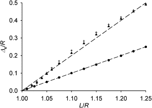
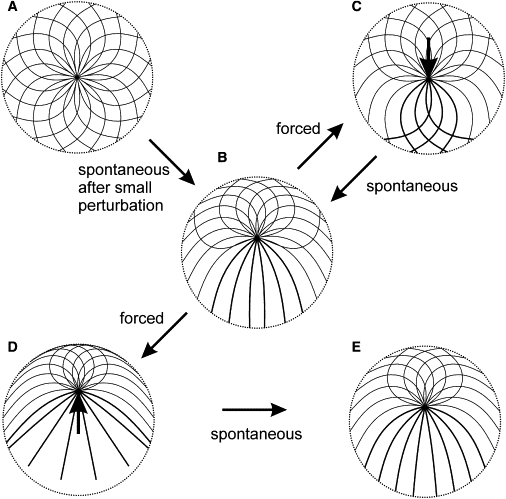
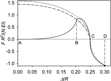
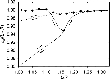
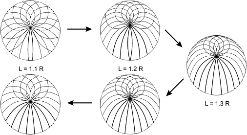
Many cell cytoskeletons include an aster of microtubules, with the centrosome serving as the focal point. The position of the centrosome within the cell is important in such directional activities as wound closure and interactions of immune cells. Here we analyzed the centrosome positioning as it is dictated by microtubule elasticity alone in a mechanical model of an intrinsically fully symmetric microtubule aster. We demonstrate that the symmetry and the central position of the centrosome are unstable. The equilibrium deviation of the centrosome from the center is approximately proportional to the difference of the microtubule length and cell radius. The proportionality coefficient is 1 in flat cells and 2 in three-dimensional cells. The loss of symmetry is irreversible, and in general, the equilibrium form of the aster exhibits memory of past perturbations. The equilibrium position of the centrosome as a function of the microtubule length exhibits hysteresis, and the history of the length variation is reflected in the aster form. These properties of the simple aster of elastic microtubules must be taken into account in the analysis of more comprehensive theoretical models, and in the design and interpretation of experiments addressing the complex process of cytoskeleton morphogenesis.


Figures

Figure 1
Figure 1
Diagram of the model for the equilibrium of a microtubule. See Table 1 for nomenclature. The bold line represents the microtubule. The circumference represents the cell boundary.
Figure 2
Figure 2
The equilibrium distance of the centrosome from the cell center. The triangles show the case of the continuous uniform spatial distribution of the unstrained directions of microtubule emanation from the centrosome in a spherical cell. The crosses show for comparison the discrete case in which 20 microtubules emanate in the directions of the vertices of a dodecahedron. The circles are the positions reached spontaneously after a small perturbation of a fully symmetric cytoskeleton in a flat round cell (in the continuous approximation as everywhere else in this article). The dashed lines have the slopes of 1 and 2 for reference. Please note that this plot starts from the (1, 0) point, and therefore the distance is approximately proportional to the excess length (length minus radius).
Figure 3
Figure 3
Conformations of idealized microtubule cytoskeletons in a flat cell. (A) The fully symmetric microtubule cytoskeleton. (B) The equilibrium which the fully symmetric cytoskeleton reaches spontaneously in response to a small perturbation. The perturbation in this example is an infinitesimal displacement of the centrosome upward in the plane of the image, which direction determines the final orientation of the centrosome. (C) The central centrosome position restored by application of an external force of 1.5 N EI/R2 as shown (see arrow). (D) The equilibrium reached from the configuration shown in panel B, when external force 1.5 N EI/R2 is applied to the centrosome in the direction away from the cell center. (E) The equilibrium reached from the configuration in panel D after removal of the external force. Curves show microtubule forms for 16 equally spaced directions of unstrained emanation from the centrosome. (There is an infinite number of such directions, and therefore microtubules, in the model, which are sampled here to show a finite number of microtubule forms in the figure.) Double-width lines show forms that are doubly represented, compared with the forms shown by the single-width lines (i.e., the double-width forms correspond to the angular density p, and the single-width forms, to p/2; see text). (Dotted circle) Cell boundary. L = 1.2 R.
Figure 4
Figure 4
Total force exerted by the microtubules on the centrosome as a function of the centrosome position in a flat cell. (Solid curve) Branch traced in the course of a continuous displacement of the centrosome away from the cell center, starting in the fully symmetric state of the cytoskeleton (from A to D). It can also be traced in the reverse direction, but only between A and B. (Dashed curve) Branch traced during forced reversed displacement of the centrosome, starting at the eccentric equilibrium position (from C to A). (Dotted curve) Branch traced during reversed displacement of the centrosome, starting at the distance of 0.3 cell radii (from D to A). L = 1.25 R. This is a plot of F as a function of Δ, with the argument and the value of the function normalized to N, R, and EI in order to show the parameter-independent behavior of the nondimensionalized model.
Figure 5
Figure 5
Equilibrium position of the centrosome as a function of microtubule length in a flat cell. (Diamonds) Position reached spontaneously after a small perturbation of a fully symmetric cytoskeleton. (Solid line) Evolution of the equilibrium position of the centrosome in the course of a continuous elongation of microtubules. The other two lines show the evolution of the equilibrium position in the course of shortening that starts from the two selected reversal points on the solid curve.
Figure 6
Figure 6
Equilibrium forms of a microtubule aster in a flat cell during elongation and subsequent shortening of the microtubules. Plotting conventions as in Fig. 3.

Similar articles

Cited by

References

    1. Bray D. Garland; New York: 2000. Cell Movements: From Molecules to Motility.
    1. Gotlieb A.I., May L.M., Kalnins V.I. Distribution of microtubule organizing centers in migrating sheets of endothelial cells. J. Cell Biol. 1981;91:589–594. - PMC - PubMed
    1. Kupfer A., Louvard D., Singer S.J. Polarization of the Golgi apparatus and the microtubule-organizing center in cultured fibroblasts at the edge of an experimental wound. Proc. Natl. Acad. Sci. USA. 1982;79:2603–2607. - PMC - PubMed
    1. Ueda M., Gräf R., Euteneuer U. Centrosome positioning and directionality of cell movements. Proc. Natl. Acad. Sci. USA. 1997;94:9674–9678. - PMC - PubMed
    1. Kupfer A., Singer S.J. Cell biology of cytotoxic and helper T cell functions: immunofluorescence microscopic studies of single cells and cell couples. Annu. Rev. Immunol. 1989;7:309–337. - PubMed

Publication types

LinkOut - more resources