. 2010 Nov 3;99(9):2936-46.
doi: 10.1016/j.bpj.2010.09.011.

Docking and fast fusion of synaptobrevin vesicles depends on the lipid compositions of the vesicle and the acceptor SNARE complex-containing target membrane

Affiliations

Docking and fast fusion of synaptobrevin vesicles depends on the lipid compositions of the vesicle and the acceptor SNARE complex-containing target membrane

Marta K Domanska et al. Biophys J. .

Erratum in

  • Biophys J. 2011 Jan 5;100(1):269

Abstract

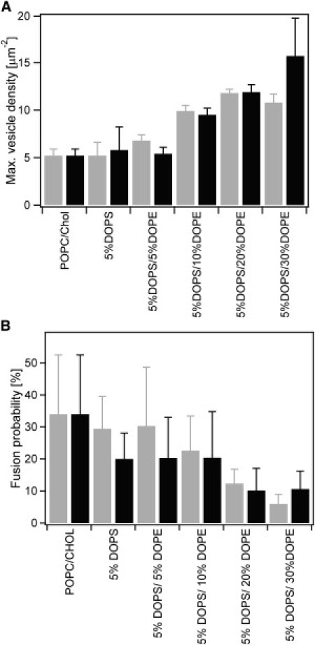
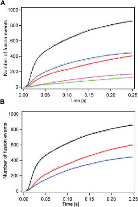
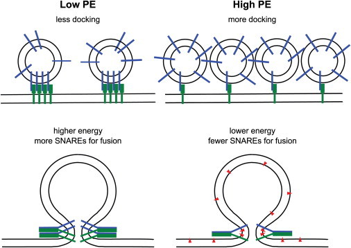
The influence of the lipid environment on docking and fusion of synaptobrevin 2 (Syb2) vesicles with target SNARE complex membranes was examined in a planar supported membrane fusion assay with high time-resolution. Previously, we showed that approximately eight SNARE complexes are required to fuse phosphatidylcholine (PC) and cholesterol model membranes in ∼20 ms. Here we present experiments, in which phosphatidylserine (PS) and phosphatidylethanolamine (PE) were added to mixtures of PC/cholesterol in different proportions in the Syb2 vesicle membranes only or in both the supported bilayers and the Syb2 vesicles. We found that PS and PE both reduce the probability of fusion and that this reduction is fully accounted for by the lipid composition in the vesicle membrane. However, the docking efficiency increases when the PE content in the vesicle (and target membrane) is increased from 0 to 30%. The fraction of fast-activating SNARE complexes decreases with increasing PE content. As few as three SNARE complexes are sufficient to support membrane fusion when at least 5% PS and 10% PE are present in both membranes or 5% and 30% PE are present in the vesicle membrane only. Despite the smaller number of required SNAREs, the SNARE activation and fusion rates are almost as fast as previously reported in reconstituted PC/cholesterol bilayers, i.e., ~10 and ~20 ms, respectively [corrected].


Figures

Figure 1
Figure 1
Lipid composition of the vesicle and target membrane affects docking and fusion of Syb2 vesicles in single vesicle to planar supported membrane fusion assay. (A) Mean final docking after saturation of reconstituted acceptor SNARE complex-containing target membranes with Syb2 vesicles. Numbers of docked vesicles per μm2 are average values of 16–18 independent experiments (different bilayers), in which the final docking densities were obtained by fitting Eq. 1. Error bars represent standard deviations of the mean final docking density between different bilayers. (B) Fusion probability calculated as the percentage of observed total fusion events over all recorded docking events. Error bars represent standard deviations from all measurements in multiple membranes of the same lipid composition. The “extracellular” leaflets in all supported target membranes were composed of POPC/Chol (4:1), whereas the “cytoplasmic” leaflets contained in addition different concentrations of DOPS and DOPE as indicated. These lipids were added to vesicle and target membranes (gray bars) or to vesicle membranes only (black bars).
Figure 2
Figure 2
Histograms of fusion delay times obtained in membranes of different lipid composition. The “extracellular” leaflets in all supported target membranes were composed of POPC/Chol (4:1), whereas the “cytoplasmic” leaflets contained in addition: (A) 5 mol % DOPS and 5 mol % DOPE, (B) 5 mol % DOPS and 10 mol % DOPE, (C) 5 mol % DOPS and 20 mol % DOPE, and (D) 5 mol % DOPS and 30 mol % DOPE. (Gray histograms on the left) Data obtained with DOPS and DOPE present in the Syb2 vesicle and planar target membrane. (Black histograms on the right) Data collected with DOPS and DOPE that is present only in Syb2 vesicle membranes.
Figure 3
Figure 3
Cumulative distribution functions of single Syb2 vesicle fusion events to acceptor SNARE-complex containing membranes of different lipid compositions. Cumulative distribution functions representing the synchronized kinetics of membrane merger were fitted with the multiparticle parallel activation model as described in the text. (Solid lines) Best-fit curves. (Squares) Experimental data. Fusion kinetics obtained in asymmetric membranes, in which DOPS and DOPE were present in Syb2 vesicle membranes only (A) and in both vesicle and target membranes (B). (Black, POPC/Chol (4:1); red, plus 5 mol % DOPS and DOPE; blue, plus 5 mol % DOPS and 10 mol % DOPE; green, plus 5 mol % DOPS and 20 mol % DOPE; magenta, plus 5 mol % DOPS and 30 mol % DOPE.)
Figure 4
Figure 4
Models explaining the effects of PE on docking and fusion. In the absence of PE, we hypothesize that SNAREs may be more clustered in cholesterol-containing target and vesicle membranes leading to fewer docking sites. The membrane-bending energy in the absence of PE is higher requiring more SNAREs for fusion. In the presence of PE, SNARE clusters may be (partially) dispersed leading to more docking sites. The inverted cone shape of PE (red triangles) facilitates curved intermediate membrane structures (including hemifusion stalk, not shown) and thus lowers the energy of fusion intermediates requiring fewer SNAREs for fusion.

References

    1. Sutton R.B., Fasshauer D., Brunger A.T. Crystal structure of a SNARE complex involved in synaptic exocytosis at 2.4 Å resolution. Nature. 1998;395:347–353. - PubMed
    1. Fasshauer D. Structural insights into the SNARE mechanism. Biochim. Biophys. Acta. 2003;1641:87–97. - PubMed
    1. Jahn R., Scheller R.H. SNAREs—engines for membrane fusion. Nat. Rev. Mol. Cell Biol. 2006;7:631–643. - PubMed
    1. Rizo J., Rosenmund C. Synaptic vesicle fusion. Nat. Struct. Mol. Biol. 2008;15:665–674. - PMC - PubMed
    1. Pobbati A.V., Stein A., Fasshauer D. N- to C-terminal SNARE complex assembly promotes rapid membrane fusion. Science. 2006;313:673–676. - PubMed

Publication types

MeSH terms