. 2010 Nov 3;99(9):3076-83.
doi: 10.1016/j.bpj.2010.09.018.

Mechanical forces regulate elastase activity and binding site availability in lung elastin

Affiliations

Mechanical forces regulate elastase activity and binding site availability in lung elastin

Rajiv Jesudason et al. Biophys J. .

Abstract

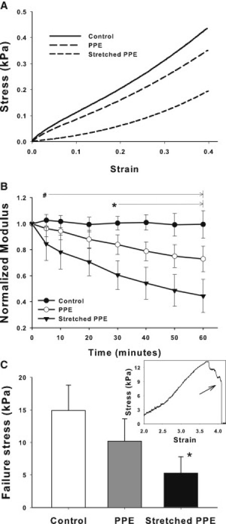
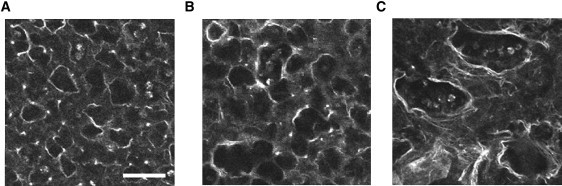
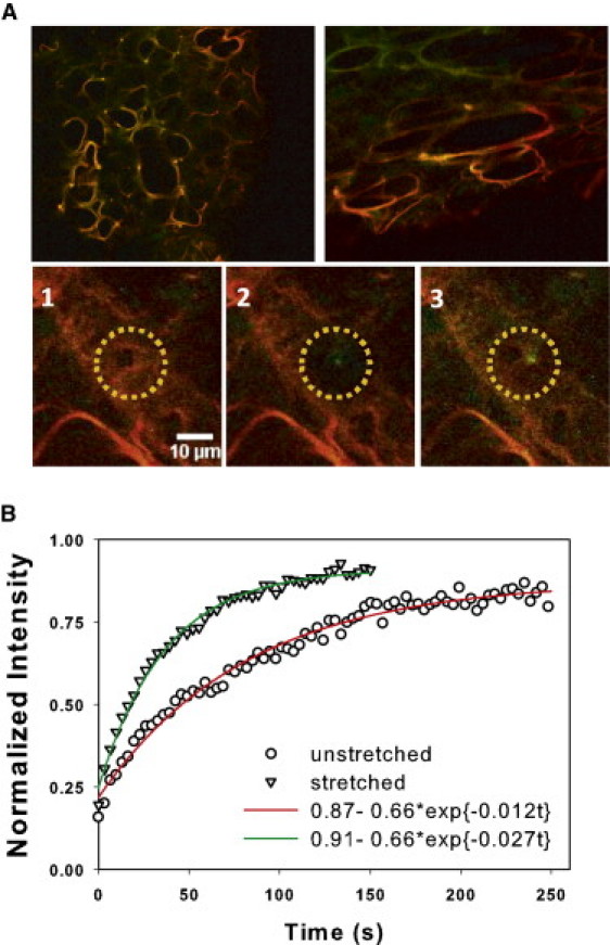
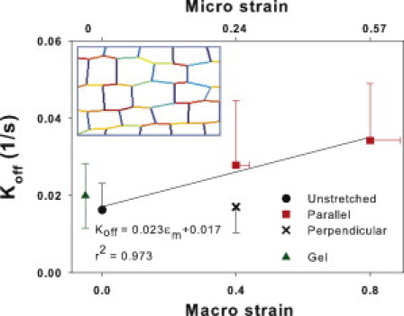
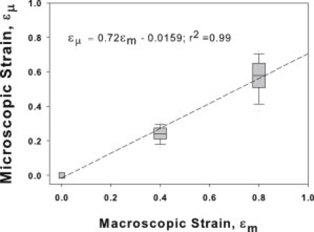
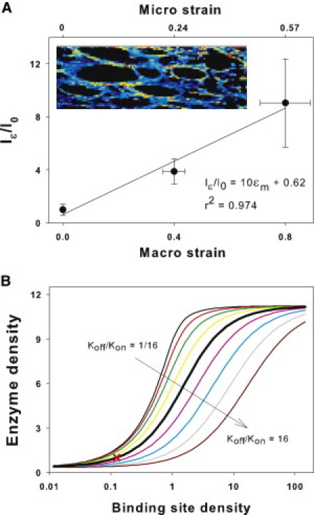
Many fundamental cellular and extracellular processes in the body are mediated by enzymes. At the single molecule level, enzyme activity is influenced by mechanical forces. However, the effects of mechanical forces on the kinetics of enzymatic reactions in complex tissues with intact extracellular matrix (ECM) have not been identified. Here we report that physiologically relevant macroscopic mechanical forces modify enzyme activity at the molecular level in the ECM of the lung parenchyma. Porcine pancreatic elastase (PPE), which binds to and digests elastin, was fluorescently conjugated (f-PPE) and fluorescent recovery after photobleach was used to evaluate the binding kinetics of f-PPE in the alveolar walls of normal mouse lungs. Fluorescent recovery after photobleach indicated that the dissociation rate constant (k(off)) for f-PPE was significantly larger in stretched than in relaxed alveolar walls with a linear relation between k(off) and macroscopic strain. Using a network model of the parenchyma, a linear relation was also found between k(off) and microscopic strain on elastin fibers. Further, the binding pattern of f-PPE suggested that binding sites on elastin unfold with strain. The increased overall reaction rate also resulted in stronger structural breakdown at the level of alveolar walls, as well as accelerated decay of stiffness and decreased failure stress of the ECM at the macroscopic scale. These results suggest an important role for the coupling between mechanical forces and enzyme activity in ECM breakdown and remodeling in development, and during diseases such as pulmonary emphysema or vascular aneurysm. Our findings may also have broader implications because in vivo, enzyme activity in nearly all cellular and extracellular processes takes place in the presence of mechanical forces.


Figures

Figure 1
Figure 1
(A) Representative stress-strain curves obtained in normal mouse lung tissue before treatment (control) and after 1 hour of porcine pancreatic elastase (PPE) digestion in the absence or presence of static 40% uniaxial stretch. (B) Means and SDs of the normalized modulus Yn as a function of time for control, PPE, and stretched PPE conditions. # denotes a statistically significant difference between the stretched PPE group and the other conditions. denotes a statistically significant difference between the PPE and the control groups. (C) Failure stress measured at the end of 1 h protocol for the three groups. denotes a statistically significant difference between the stretched PPE and the control groups. The inset shows an example failure stress-strain curve and the arrow indicates the failure stress.
Figure 2
Figure 2
Autofluorescent images showing alveolar structure at 1 h taken in the unstretched condition of the tissue strips. The left, middle, and right panels show images in the control, the PPE digested, and the PPE digested in the presence of 40% static uniaxial strain, respectively. Note the significantly increased aspect ratio after digestion under stretch. Scale bar denotes 50 μm.
Figure 3
Figure 3
(A) Top row shows a control sample (left) after equilibration and a stretched sample (right) before full equilibration with f-PPE. For the stretched sample, strain is in the horizontal direction. (Green) Autofluorescence; (red) f-PPE signal. (Bottom row) Three images of a region before FRAP (left), after photobleaching (middle), and after near full recovery (right). (B) Example of normalized FRAP curves from a relaxed (circles) and a stretched (triangles) alveolar wall. The fluorescent intensity was normalized to 1 at time 0 corresponding to the average intensity of the bleached region (yellow circle) on the left panel in A. (Solid lines) Single exponential model fits.
Figure 4
Figure 4
Means and SDs of estimated dissociation rates, koff, as a function of uniaxial macroscopic strain ɛm (bottom x axis) in unstretched samples (solid circle) and at ɛm of 0.4 and 0.8 (squares) measured on walls that had a direction parallel to macroscopic strain. At em = 0.4, koff was also measured along walls that were perpendicular to the macroscopic strain (cross). Inset shows a region of a nonlinear elastic network at em = 0.4 that was used to predict the average microscopic strain ɛμ (top horizontal axis) on the walls parallel to macroscopic strain. The shades (colors in the Supporting Material) represent local microscopic strain darker (red) denoting higher strains. Note the large variability among horizontal segments. The SD of ɛμ is plotted as horizontal error bars. The solid line shows the linear regression. The mean and SD of koff measured in pure elastin hydrogel is also shown (triangle).
Figure 5
Figure 5
Microscopic strain as a function of uniaxial macroscopic strain in the hexagonal network model of the lung tissue. (Line) Linear regression.
Figure 6
Figure 6
(A) Fluorescent intensity on alveolar walls parallel to macroscopic strain (Iɛ) normalized to mean intensity in the unstretched samples (I0) as a function of macroscopic and microscopic strains, as described in Fig. 3's legend. (Inset) Zoom into an alveolar region with brighter shades (shown in colors in the Supporting Material) representing high f-PPE intensity normalized by the autofluorescent intensity. The shades (colors) demonstrate a strong anisotropy as well as heterogeneity in labeling along parallel wall segments. (B) Sensitivity of model-based total enzyme concentration on a fiber, as a surrogate for fluorescent intensity, to variations in binding site density B0 for a range of binding on rates kon based on Eq. 4. (Thick black line) Baseline model for which B0 = 1/8 corresponds to unit enzyme concentration (cross).

Similar articles

Cited by

References

    1. Alberts B. Garland Science; New York: 2008. Molecular Biology of the Cell.
    1. Ayala Y.M., Di Cera E. A simple method for the determination of individual rate constants for substrate hydrolysis by serine proteases. Protein Sci. 2000;9:1589–1593. - PMC - PubMed
    1. Joubert F., Fales H.M., Balaban R.S. NADH enzyme-dependent fluorescence recovery after photobleaching (ED-FRAP): applications to enzyme and mitochondrial reaction kinetics, in vitro. Biophys. J. 2004;86:629–645. - PMC - PubMed
    1. Mecham R.P., Broekelmann T.J., Senior R.M. Elastin degradation by matrix metalloproteinases. Cleavage site specificity and mechanisms of elastolysis. J. Biol. Chem. 1997;272:18071–18076. - PubMed
    1. Khan S., Sheetz M.P. Force effects on biochemical kinetics. Annu. Rev. Biochem. 1997;66:785–805. - PubMed

Publication types

MeSH terms

LinkOut - more resources