

Substance P induces the reversible formation of varicosities in the dendrites of rat brainstem neurons
- PMID: 21044613
- PMCID: PMC3014421
- DOI: 10.1016/j.brainres.2010.10.091
Substance P induces the reversible formation of varicosities in the dendrites of rat brainstem neurons
Abstract
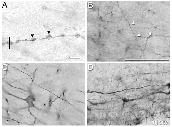
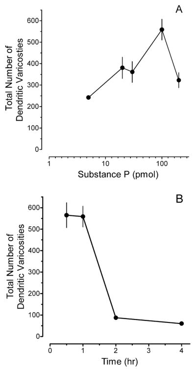
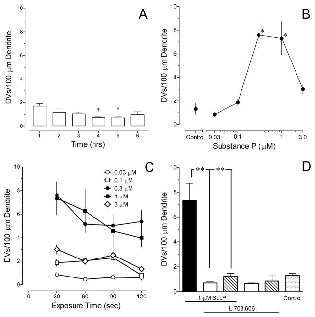
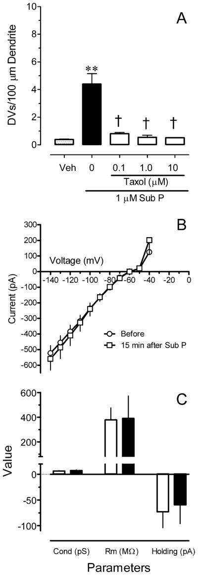
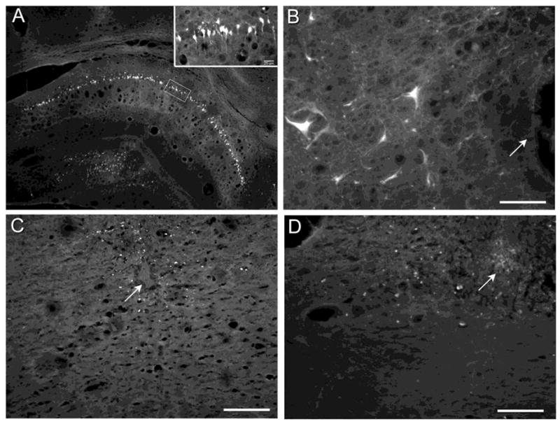
This study investigated the ability of substance P (Sub P) to induce dendritic varicosities (DVs) or beads in neurons of the rostral ventromedial medulla (RVM) of the rat. Microinjection of 5-200 pmol Sub P in the RVM produced a concentration-dependent increase in the number of DVs in distal dendrites of RVM neurons that were immunoreactive for the neurokinin-1 receptor, but not serotonin. The effect was reversible, as DVs were essentially absent 2 and 4h after microinjection. Fluoro-Jade B labeled neurons were not evident in the RVM 4 days after microinjection of Sub P, although such neurons were present 4 days after microinjection of a neurotoxic dose of kainate. Bath application of Sub P to brainstem slices for a period as brief as 30s also produced DVs in neurokinin-1 immunoreactive RVM neurons. Prior exposure to L-703606 prevented the formation of DVs by Sub P, implicating the neurokinin-1 receptor, a Gq type of G protein coupled receptor, in the formation of DVs by Sub P. Finally, stabilization of microtubules by prior exposure to taxol also prevented the formation of DVs, consistent with the idea that increases in intracellular Ca(2+) lead to the formation of DVs secondary to a disruption of the linear arrays of microtubules in dendrites. These data establish a mechanistic basis for the formation of DVs by Sub P and support further studies to test the hypothesis that the formation of DVs is a morphological mechanism by which neurons can regulate their responses to inhibitory or excitatory inputs.
Copyright © 2010 Elsevier B.V. All rights reserved.
Figures
Similar articles
-
Loss of neurons in rostral ventromedial medulla that express neurokinin-1 receptors decreases the development of hyperalgesia.Neuroscience. 2013 Oct 10;250:151-65. doi: 10.1016/j.neuroscience.2013.06.057. Epub 2013 Jul 3. Neuroscience. 2013. PMID: 23831426 Free PMC article.
-
Increased neuronal expression of neurokinin-1 receptor and stimulus-evoked internalization of the receptor in the rostral ventromedial medulla of the rat after peripheral inflammatory injury.J Comp Neurol. 2014 Sep 1;522(13):3037-51. doi: 10.1002/cne.23564. J Comp Neurol. 2014. PMID: 24639151 Free PMC article.
-
Substance P enhances excitatory synaptic transmission on spinally projecting neurons in the rostral ventromedial medulla after inflammatory injury.J Neurophysiol. 2009 Aug;102(2):1139-51. doi: 10.1152/jn.91337.2008. Epub 2009 Jun 3. J Neurophysiol. 2009. PMID: 19494188 Free PMC article.
-
Effects of neurokinin-1 receptor agonism and antagonism in the rostral ventromedial medulla of rats with acute or persistent inflammatory nociception.Neuroscience. 2010 Feb 3;165(3):902-13. doi: 10.1016/j.neuroscience.2009.10.064. Epub 2009 Nov 3. Neuroscience. 2010. PMID: 19892001 Free PMC article.
-
Spinal cord mechanisms mediating behavioral hyperalgesia induced by neurokinin-1 tachykinin receptor activation in the rostral ventromedial medulla.Neuroscience. 2010 Dec 29;171(4):1341-56. doi: 10.1016/j.neuroscience.2010.09.040. Epub 2010 Oct 1. Neuroscience. 2010. PMID: 20888891 Free PMC article.
Cited by
-
Hyperalgesia and sensitization of dorsal horn neurons following activation of NK-1 receptors in the rostral ventromedial medulla.J Neurophysiol. 2017 Nov 1;118(5):2727-2744. doi: 10.1152/jn.00478.2017. Epub 2017 Aug 9. J Neurophysiol. 2017. PMID: 28794197 Free PMC article.
-
Loss of neurons in rostral ventromedial medulla that express neurokinin-1 receptors decreases the development of hyperalgesia.Neuroscience. 2013 Oct 10;250:151-65. doi: 10.1016/j.neuroscience.2013.06.057. Epub 2013 Jul 3. Neuroscience. 2013. PMID: 23831426 Free PMC article.
-
Changes in the disposition of substance P in the rostral ventromedial medulla after inflammatory injury in the rat.Neuroscience. 2016 Mar 11;317:1-11. doi: 10.1016/j.neuroscience.2015.12.054. Epub 2016 Jan 4. Neuroscience. 2016. PMID: 26762802 Free PMC article.
-
Increased neuronal expression of neurokinin-1 receptor and stimulus-evoked internalization of the receptor in the rostral ventromedial medulla of the rat after peripheral inflammatory injury.J Comp Neurol. 2014 Sep 1;522(13):3037-51. doi: 10.1002/cne.23564. J Comp Neurol. 2014. PMID: 24639151 Free PMC article.
References
-
- Al-Noori S, Swann JW. A role for sodium and chloride in kainic acid-induced beading of inhibitory interneuron dendrites. Neuroscience. 2000;101:337–348. - PubMed
-
- Carrera I, Molist P, Anadon R, Rodriguez-Moldes I. Development of the serotoninergic system in the central nervous system of a shark, the lesser spotted dogfish Scyliorhinus canicula. J Comp Neurol. 2008;511:804–831. - PubMed
Publication types
MeSH terms
Substances
Grants and funding
LinkOut - more resources
Full Text Sources
Miscellaneous
