

Comparative analysis of combination kanamycin-furosemide versus kanamycin alone in the mouse cochlea
- PMID: 21044672
- PMCID: PMC4519356
- DOI: 10.1016/j.heares.2010.10.011
Comparative analysis of combination kanamycin-furosemide versus kanamycin alone in the mouse cochlea
Abstract
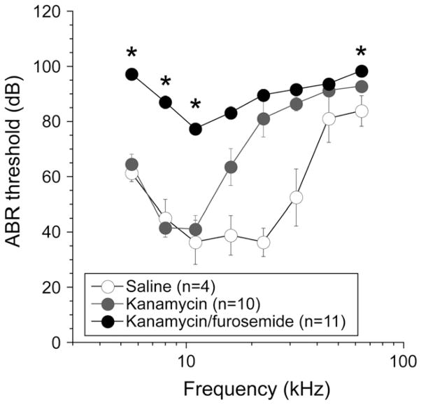
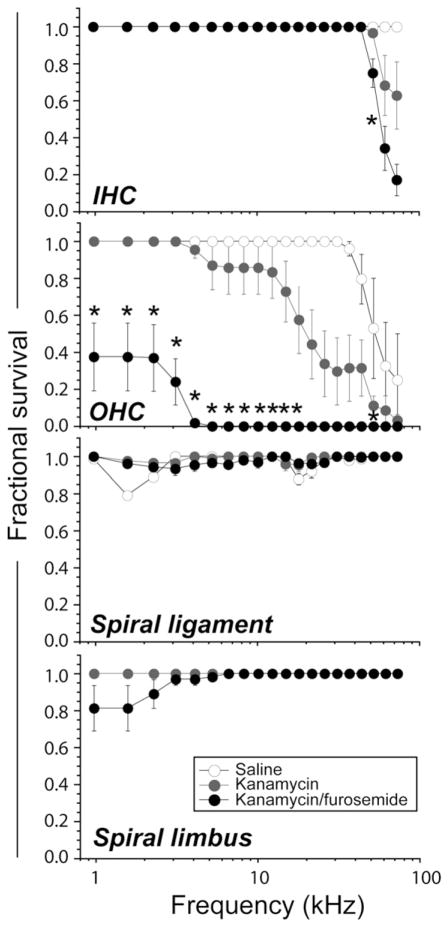
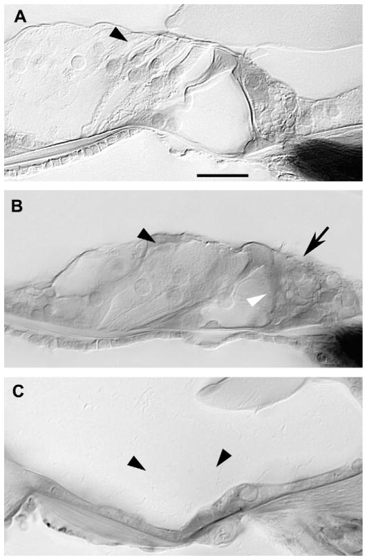
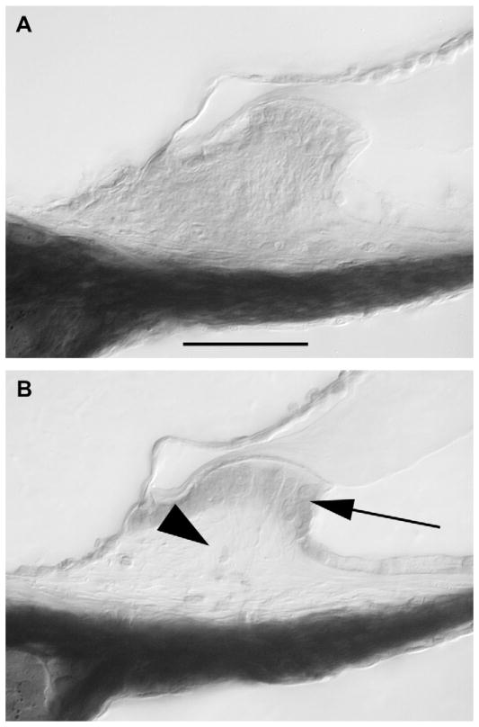
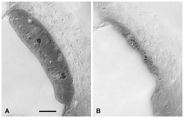
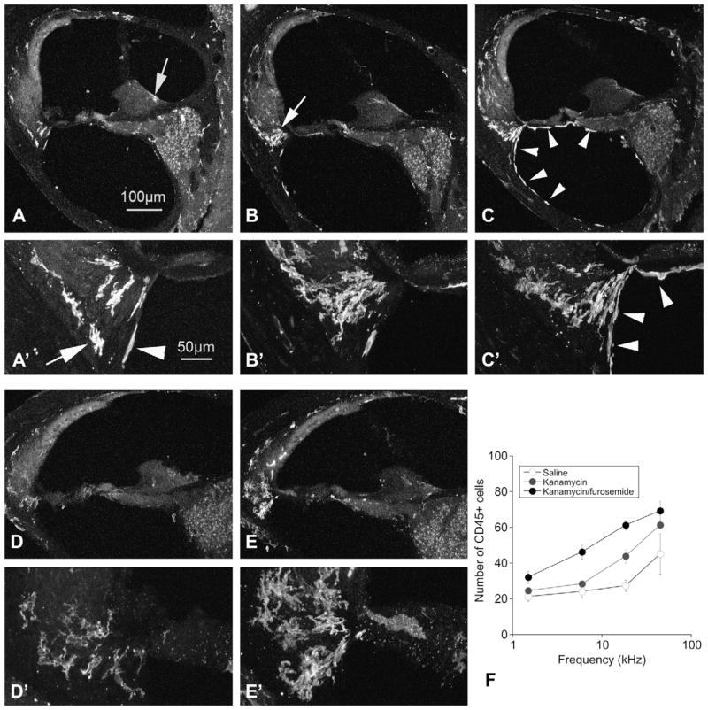
Combinations of aminoglycosides and loop diuretics have been known to have a synergistic effect in ototoxic injury. Because murine hair cells are relatively resistant to ototoxicity compared to those of other mammals, investigators have turned to combination therapies to create ototoxic lesions in the mouse inner ear. In this paper, we perform a systematic comparison of hearing thresholds, hair cell damage and monocyte migration into the mouse cochlea after kanamycin versus combined kanamycin/furosemide and explore the pathophysiology of enhanced hair cell loss in aminoglycoside ototoxicity in the presence of loop diuretic. Combined kanamycin-furosemide resulted in elevation of threshold not only in the high frequencies, but across all frequencies with more extensive loss of outer hair cells when compared to kanamycin alone. The stria vascularis was severely atrophied and stellate cells in the spiral limbus were missing in kanamycin-furosemide exposed mice while these changes were not observed in mice receiving kanamycin alone. Monocytes and macrophages were recruited in large numbers to the spiral ligament and spiral ganglion in these mice. Combination therapy resulted in a greater number of macrophages in total, and many more macrophages were present further apically when compared to mice given kanamycin alone. Combined kanamycin-furosemide provides an effective method of addressing the relative resistance to ototoxicity that is observed in most mouse strains. As the mouse becomes increasingly more common in studies of hearing loss, and combination therapies gain popularity, recognition of the overall effects of combined aminoglycoside-loop diuretic therapy will be critical to interpretation of the interventions that follow.
Copyright © 2010. Published by Elsevier B.V.
Figures
References
-
- Abrashkin KA, Izumikawa M, Miyazawa T, Wang CH, Crumling MA, Swiderski DL, Beyer LA, Gong TW, Raphael Y. The fate of outer hair cells after acoustic or ototoxic insults. Hear Res. 2006;218:20–29. - PubMed
-
- Agterberg MJ, Versnel H, de Groot JC, Smoorenburg GF, Albers FW, Klis SF. Morphological changes in spiral ganglion cells after intracochlear application of brain-derived neurotrophic factor in deafened guinea pigs. Hear Res. 2008;244:25–34. - PubMed
-
- Alam SA, Ikeda K, Kawase T, Kikuchi T, Katori Y, Watanabe K, Takasaka T. Acute effects of combined administration of kanamycin and furosemide on the stria vascularis studied by distortion product otoacoustic emission and transmission electron microscopy. Tohoku J Exp Med. 1998;186:79–86. - PubMed
-
- Arnold W, Nadol JB, Jr, Weidauer H. Ultrastructural histopathology in a case of human ototoxicity due to loop diuretics. Acta Otolaryngol. 1981;91:399–414. - PubMed
-
- Asakuma S, Snow JB., Jr Effects of furosemide and ethacrynic acid on the endocochlear direct current potential in normal and kanamycin sulfate-treated guinea pigs. Otolaryngol Head Neck Surg. 1980;88:188–193. - PubMed
Publication types
MeSH terms
Substances
Grants and funding
LinkOut - more resources
Full Text Sources
Research Materials
