Diffusion and light-dependent compartmentalization of transducin
- PMID: 21044685
- PMCID: PMC3018668
- DOI: 10.1016/j.mcn.2010.10.006
Diffusion and light-dependent compartmentalization of transducin
Abstract
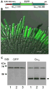
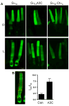
Diffusion and light-dependent compartmentalization of transducin are essential for phototransduction and light adaptation of rod photoreceptors. Here, transgenic Xenopus laevis models were designed to probe the roles of transducin/rhodopsin interactions and lipid modifications in transducin compartmentalization, membrane mobility, and light-induced translocation. Localization and diffusion of EGFP-fused rod transducin-α subunit (Gα(t1)), mutant Gα(t1) that is predicted to be N-acylated and S-palmitoylated (Gα(t1)A3C), and mutant Gα(t1) uncoupled from light-activated rhodopsin (Gα(t1)-Ctα(s)), were examined by EGFP-fluorescence imaging and fluorescence recovery after photobleaching (FRAP). Similar to Gα(t1), Gα(t1)A3C and Gα(t1)-Ctα(s) were correctly targeted to the rod outer segments in the dark, however the light-dependent translocation of both mutants was markedly impaired. Our analysis revealed a moderate acceleration of the lateral diffusion for the activated Gα(t1) consistent with the diffusion of the separated Gα(t1)GTP and Gβ(1)γ(1) on the membrane surface. Unexpectedly, the kinetics of longitudinal diffusion were comparable for Gα(t1)GTP with a single lipid anchor and heterotrimeric Gα(t1)β(1)γ(1) or Gα(t1)-Ctα(s)β(1)γ(1) with two lipid modifications. This contrasted the lack of the longitudinal diffusion of the Gα(t1)A3C mutant apparently caused by its stable two lipid attachment to the membrane and suggests the existence of a mechanism that facilitates axial diffusion of Gα(t1)β(1)γ(1).
Copyright © 2010 Elsevier Inc. All rights reserved.
Figures


Similar articles
-
Interaction of transducin with uncoordinated 119 protein (UNC119): implications for the model of transducin trafficking in rod photoreceptors.J Biol Chem. 2011 Aug 19;286(33):28954-28962. doi: 10.1074/jbc.M111.268821. Epub 2011 Jun 28. J Biol Chem. 2011. PMID: 21712387 Free PMC article.
-
Functional comparison of rod and cone Gα(t) on the regulation of light sensitivity.J Biol Chem. 2013 Feb 22;288(8):5257-67. doi: 10.1074/jbc.M112.430058. Epub 2013 Jan 3. J Biol Chem. 2013. PMID: 23288843 Free PMC article.
-
Rod phosphodiesterase-6 PDE6A and PDE6B subunits are enzymatically equivalent.J Biol Chem. 2010 Dec 17;285(51):39828-34. doi: 10.1074/jbc.M110.170068. Epub 2010 Oct 12. J Biol Chem. 2010. PMID: 20940301 Free PMC article.
-
Light-dependent compartmentalization of transducin in rod photoreceptors.Mol Neurobiol. 2008 Feb;37(1):44-51. doi: 10.1007/s12035-008-8015-2. Epub 2008 Apr 19. Mol Neurobiol. 2008. PMID: 18425604 Review.
-
A physiological role for the supramolecular organization of rhodopsin and transducin in rod photoreceptors.FEBS Lett. 2013 Jun 27;587(13):2060-6. doi: 10.1016/j.febslet.2013.05.017. Epub 2013 May 16. FEBS Lett. 2013. PMID: 23684654 Review.
Cited by
-
Frmpd1 Facilitates Trafficking of G-Protein Transducin and Modulates Synaptic Function in Rod Photoreceptors of Mammalian Retina.eNeuro. 2022 Oct 17;9(5):ENEURO.0348-22.2022. doi: 10.1523/ENEURO.0348-22.2022. Print 2022 Sep-Oct. eNeuro. 2022. PMID: 36180221 Free PMC article.
-
Ift172 conditional knock-out mice exhibit rapid retinal degeneration and protein trafficking defects.Hum Mol Genet. 2018 Jun 1;27(11):2012-2024. doi: 10.1093/hmg/ddy109. Hum Mol Genet. 2018. PMID: 29659833 Free PMC article.
-
In vivo optophysiology reveals that G-protein activation triggers osmotic swelling and increased light scattering of rod photoreceptors.Proc Natl Acad Sci U S A. 2017 Apr 4;114(14):E2937-E2946. doi: 10.1073/pnas.1620572114. Epub 2017 Mar 20. Proc Natl Acad Sci U S A. 2017. PMID: 28320964 Free PMC article.
-
Protein sorting, targeting and trafficking in photoreceptor cells.Prog Retin Eye Res. 2013 Sep;36:24-51. doi: 10.1016/j.preteyeres.2013.03.002. Epub 2013 Apr 3. Prog Retin Eye Res. 2013. PMID: 23562855 Free PMC article. Review.
-
Cul3-Klhl18 ubiquitin ligase modulates rod transducin translocation during light-dark adaptation.EMBO J. 2019 Dec 2;38(23):e101409. doi: 10.15252/embj.2018101409. Epub 2019 Nov 7. EMBO J. 2019. PMID: 31696965 Free PMC article.
References
-
- Artemyev NO. Light-dependent compartmentalization of transducin in rod photoreceptors. Mol Neurobiol. 2008;37:44–51. - PubMed
-
- Bigay J, Faurobert E, Franco M, Chabre M. Roles of lipid modifications of transducin subunits in their GDP-dependent association and membrane binding. Biochemistry. 1994;33:14081–14090. - PubMed
-
- Bourne HR. How receptors talk to trimeric G proteins. Curr Opin Cell Biol. 1997;9:134–142. - PubMed
-
- Burns ME, Arshavsky VY. Beyond counting photons: trials and trends in vertebrate visual transduction. Neuron. 2005;48:387–401. - PubMed
Publication types
MeSH terms
Substances
Grants and funding
LinkOut - more resources
Full Text Sources
Miscellaneous

