. 2010 Nov 16;107(46):19915-20.
doi: 10.1073/pnas.1009023107. Epub 2010 Nov 2.

Beta-N-acetylglucosamine (O-GlcNAc) is part of the histone code

Affiliations

Beta-N-acetylglucosamine (O-GlcNAc) is part of the histone code

Kaoru Sakabe et al. Proc Natl Acad Sci U S A. .

Abstract

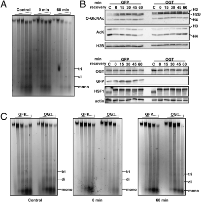
Dynamic posttranslational modification of serine and threonine residues of nucleocytoplasmic proteins by β-N-acetylglucosamine (O-GlcNAc) is a regulator of cellular processes such as transcription, signaling, and protein-protein interactions. Like phosphorylation, O-GlcNAc cycles in response to a wide variety of stimuli. Although cycling of O-GlcNAc is catalyzed by only two highly conserved enzymes, O-GlcNAc transferase (OGT), which adds the sugar, and β-N-acetylglucosaminidase (O-GlcNAcase), which hydrolyzes it, the targeting of these enzymes is highly specific and is controlled by myriad interacting subunits. Here, we demonstrate by multiple specific immunological and enzymatic approaches that histones, the proteins that package DNA within the nucleus, are O-GlcNAcylated in vivo. Histones also are substrates for OGT in vitro. We identify O-GlcNAc sites on histones H2A, H2B, and H4 using mass spectrometry. Finally, we show that histone O-GlcNAcylation changes during mitosis and with heat shock. Taken together, these data show that O-GlcNAc cycles dynamically on histones and can be considered part of the histone code.


Conflict of interest statement

The authors declare no conflict of interest.

Figures

Fig. 1.
Fig. 1.
Histones are modified with O-GlcNAc. (A) Acid-extracted histones were labeled using the Click-IT kit (Invitrogen). Briefly, GlcNAc moieties were labeled with an azido-modified Gal using mGalT1. The azido sugar was reacted further with a biotin alkyne, thereby tagging O-GlcNAc residues with biotin. Blots then were probed with streptavidin-HRP (Strep-HRP) to reveal O-GlcNAcylated proteins and stained with colloidal gold for total protein. (B) O-GlcNAc on histones was labeled with 3H-Gal using GalT and UDP-[3H]-Gal. The upper gel represents the total protein, and the lower gel is the autoradiograph. (C) Natively purified histones were probed with two separate antibodies that recognize O-GlcNAc, RL2 (Left) and 110.6 (Right). For specificity, 1 M GlcNAc competition blots were included. Blots also were probed with histone H2B and were stained with colloidal gold for protein loading. (D) FLAG-tagged histones were immunoprecipitated from transfected HeLa cells and probed for O-GlcNAc (RL2), FLAG, and H2B. Asterisks indicate the FLAG-tagged histones. (E) Acid-extracted histones from asynchronously growing HeLa cells (Upper) and HeLa cells treated with sodium butyrate (Lower) were separated in two dimensions. The first dimension was based on charge (AU), and the second dimension was based on molecular weight (MW). Blots also were probed with H2B for loading. (F) Acid-extracted histones were resolved as in E and probed for O-GlcNAc with or without 1 M GlcNAc for specificity.
Fig. 2.
Fig. 2.
Identification of O-GlcNAcylation sites on histones. (A) MS/MS spectrum of the precursor ion ([M+2H]2+ 658.62) carrying a DTT moiety identifying Ser47 of histone H4 as O-GlcNAcylated. (B) MS/MS spectrum of the precursor ion ([M+2H]2+ 708.5) carrying a DTT moiety identifying Ser36 of histone H2B as O-GlcNAcylated. (C) MS/MS spectrum of precursor ion ([M+2H]2+ 1034.2) identifying Thr101 as the O-GlcNAc site on histone H2A. Asterisks indicate ions containing the DTT tag. A, Ala; E, Glu; G, Gly; I, Ile; K, Lys; L, Leu; P, Pro; Q, Gln; R, Arg; S, Ser; T, Thr; V, Val; Y, Tyr.
Fig. 3.
Fig. 3.
Validation of mapped O-GlcNAc sites. (A) Core nucleosomal particle is colored in nude. (For simplicity, histone tails are not shown.) DNA is in green. H4 sites are labeled in blue, H2B sites in red, and H2A sites in green. (B) Ser47 of modified H4 is shown in blue as a stick diagram. The tail of H4 is shown in red as a ribbon. (C) Thr101 of H2A is shown in green as a stick diagram, H4 is shown in red, H3 is shown in pink, and H2A is shown in blue. The α2 and α3 domains of H3 and H4 are labeled. (D) Ser36 of H2B is shown in red in a stick diagram. The histone tail of H2B is shown in blue. To verify site-mapping data, HeLa cells were transfected with FLAG-tagged constructs for wild type and corresponding Ser/Thr-to-Ala mutations for H2A (E), H2B (F), and H4 (G). Blots were probed for O-GlcNAc (RL2) and FLAG.
Fig. 4.
Fig. 4.
Total histone O-GlcNAcylation changes with heat shock. (A) HeLa cells were heat shocked for 1 h at 45 °C and allowed to recover for the indicated times. Histones were probed for O-GlcNAc (RL2) and for H2B. (Left) Densitometric analysis of relative O-GlcNAc levels on histones normalized to histone H2B. (Right) Soluble extracts were probed for HSF1 and actin as controls. (B) In nucleo OGT assays were performed. The upper panel is the autoradiograph, which shows that histones H2A and H4 are major targets for OGT in this assay. The lower panel is the Coomassie G250 stain for total protein. (C) Densitometry of 3H-GlcNAc levels normalized to the amount of histones. (D) OGT was immunoprecipitated, and activity assays were performed using CK2 peptide as substrate. Activity is presented as pmol GlcNAc released per mg OGT immunoprecipitated min assay.
Fig. 5.
Fig. 5.
OGT overexpression affects chromatin condensation. (A) Chromatin-sensitivity assays were performed. Mono-, di-, and trinucleosomes are marked. Digestions with MNase were performed for 0, 2, 5, 10, and 15 min. (B) Histones from cells overexpressing GFP or OGT exposed to 1 h heat shock at 45 °C and allowed to recover at 37 °C as indicated were probed for O-GlcNAc and acetylated Lys (AcK). Histones H3, H2B, and H4 are indicated. Soluble extract also was probed for OGT, GFP, HSF1, and actin. (C) Chromatin-sensitivity assays were performed on cells overexpressing GFP or OGT from control cells or cells that had been heat shocked for 1 h at 45 °C and allowed to recover for 0 or 60 min at 37 °C. Digestions with MNase were performed as above.

Comment in

References

    1. Torres CR, Hart GW. Topography and polypeptide distribution of terminal N-acetylglucosamine residues on the surfaces of intact lymphocytes. Evidence for O-linked GlcNAc. J Biol Chem. 1984;259:3308–3317. - PubMed
    1. Wang Z, et al. Enrichment and site-mapping of O-linked N-acetylglucosamine by a combination of chemical/enzymatic tagging, photochemical cleavage, and electron transfer dissociation (ETD) mass spectrometry. Mol Cell Proteomics. 2009;9:153–160. - PMC - PubMed
    1. Hart GW, Housley MP, Slawson C. Cycling of O-linked beta-N-acetylglucosamine on nucleocytoplasmic proteins. Nature. 2007;446:1017–1022. - PubMed
    1. Wang Z, Gucek M, Hart GW. Cross-talk between GlcNAcylation and phosphorylation: Site-specific phosphorylation dynamics in response to globally elevated O-GlcNAc. Proc Natl Acad Sci USA. 2008;105:13793–13798. - PMC - PubMed
    1. Dias WB, Hart GW. O-GlcNAc modification in diabetes and Alzheimer's disease. Mol Biosyst. 2007;3:766–772. - PubMed

Publication types

LinkOut - more resources