. 2010 Nov 1;66(Pt 11):1503-7.
doi: 10.1107/S1744309110036535. Epub 2010 Oct 29.

Purification, crystallization and preliminary X-ray diffraction analysis of the carbohydrate-binding region of the Streptococcus gordonii adhesin GspB

Affiliations

Purification, crystallization and preliminary X-ray diffraction analysis of the carbohydrate-binding region of the Streptococcus gordonii adhesin GspB

Tasia M Pyburn et al. Acta Crystallogr Sect F Struct Biol Cryst Commun. .

Abstract

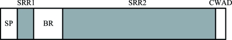
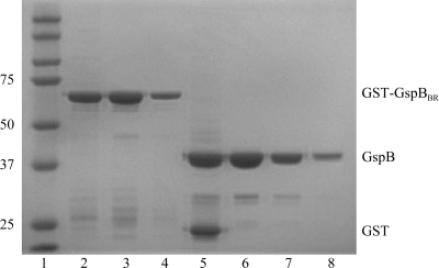
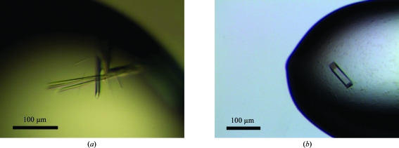
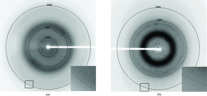
The carbohydrate-binding region of the bacterial adhesin GspB from Streptococcus gordonii strain M99 (GspB(BR)) was expressed in Escherichia coli and purified using affinity and size-exclusion chromatography. Separate sparse-matrix screening of GspB(BR) buffered in either 20 mM Tris pH 7.4 or 20 mM HEPES pH 7.5 resulted in different crystallographic behavior such that different precipitants, salts and additives supported crystallization of GspB(BR) in each buffer. While both sets of conditions supported crystal growth in space group P2(1)2(1)2(1), the crystals had distinct unit-cell parameters of a = 33.3, b = 86.7, c = 117.9 Å for crystal form 1 and a = 34.6, b = 98.3, c = 99.0 Å for crystal form 2. Additive screening improved the crystals grown in both conditions such that diffraction extended to beyond 2 Å resolution. A complete data set has been collected to 1.3 Å resolution with an overall R(merge) value of 0.04 and an R(merge) value of 0.33 in the highest resolution shell.


Figures

Figure 1
Figure 1
Overall architecture of GspB. GspB is comprised of a signal peptide (SP), a short serine-rich region (SRR1), a unique basic region (BR) that is responsible for carbohydrate binding, a second, longer, serine-rich region (SRR2) and a cell-wall-anchoring domain (CWAD). A recently published structural study of the Fap1 adhesin from S. parasanguinis identified the structural elements of both the serine-rich repeats and the unique region in that protein (Ramboarina et al., 2010 ▶). The repeat region of GspB is likely to form a super-helical fibril like that observed in the repeat region of Fap1.
Figure 2
Figure 2
Purification of GspBBR for crystallization. The gel shows two separate concentrations of the GST-GspBBR fusion protein after affinity purification and two concentrations of purified isolated GspBBR(246–601) after size-exclusion chromatography. Lane 1, Kaleidoscope Ladder (Bio-Rad; labeled in kDa); lanes 2 and 3, 4 µg GST-GspBBR; lane 4, 1 µg GST-GspBBR; lane 5, 4 µg of GST and GspBBR after factor Xa cleavage; lane 6, 4 µg GspBBR after size-exclusion chromatography; lane 7, 1 µg GspBBR after size-exclusion chromatography; lane 8, dissolved crystals grown in 26% Jeffamine-ED 2001 and 0.05 M HEPES pH 7.5.
Figure 3
Figure 3
Crystals of GspBBR from S. gordonii strain M99 grown using the hanging-drop vapor-diffusion method. (a) Crystals of GspBBR grown from 25% PEG 3350, 0.15 M ammonium acetate, 0.1 M HEPES pH 7.5 and 10 mM spermidine. (b) Crystals of GspBBR grown from 34% Jeffamine ED-2001, 0.15 M KCl and 0.1 M HEPES pH 7.5.
Figure 4
Figure 4
Typical diffraction for each of the crystal forms. (a) Diffraction image for crystals grown using PEG 3350 as the precipitant. This diffraction image was collected on the SSRL 11-1 beamline and diffraction to 1.8 Å resolution can be observed. (b) Typical diffraction image for crystals grown using Jeffamine ED-2001 as the precipitant. This diffraction image was collected on the LS-CAT ID-21-G beamline and diffraction to 1.3 Å resolution can be observed.

References

    1. Bensing, B. A. & Sullam, P. M. (2002). Mol. Microbiol.44, 1081–1094. - PubMed
    1. Collaborative Computational Project, Number 4 (1994). Acta Cryst. D50, 760–763. - PubMed
    1. Cowtan, K. (1994). Jnt CCP4/ESF–EACBM Newsl. Protein Crystallogr.31, 34–38.
    1. La Fortelle, E. de & Bricogne, G. (1997). Methods Enzymol.276, 472–494. - PubMed
    1. Matthews, B. W. (1968). J. Mol. Biol.33, 491–497. - PubMed

Publication types

MeSH terms

Substances