. 2011 Feb;19(2):318-25.
doi: 10.1038/mt.2010.236. Epub 2010 Nov 2.

Surface characteristics of nanoparticles determine their intracellular fate in and processing by human blood-brain barrier endothelial cells in vitro

Affiliations

Surface characteristics of nanoparticles determine their intracellular fate in and processing by human blood-brain barrier endothelial cells in vitro

Julia V Georgieva et al. Mol Ther. 2011 Feb.

Abstract

A polarized layer of endothelial cells that comprises the blood-brain barrier (BBB) precludes access of systemically administered medicines to brain tissue. Consequently, there is a need for drug delivery vehicles that mediate transendothelial transport of such medicines. Endothelial cells use a variety of endocytotic pathways for the internalization of exogenous materials, including clathrin-mediated endocytosis, caveolar endocytosis, and macropinocytosis. The different modes of endocytosis result in the delivery of endocytosed material to distinctive intracellular compartments and therewith correlated differential processing. To obtain insight into the properties of drug delivery vehicles that direct their intracellular processing in brain endothelial cells, we investigated the intracellular processing of fixed-size nanoparticles in an in vitro BBB model as a function of distinct nanoparticle surface modifications. Caveolar endocytosis, adsorptive-mediated endocytosis, and receptor-mediated endocytosis were promoted by the use of uncoated 500-nm particles, attachment of the cationic polymer polyethyleneimine (PEI), and attachment of prion proteins, respectively. We demonstrate that surface modifications of nanoparticles, including charge and protein ligands, affect their mode of internalization by brain endothelial cells and thereby their subcellular fate and transcytotic potential.


Figures

Figure 1
Figure 1
Uptake of nanoparticles by hCMEC/D3 cells. hCMEC/D3 cells were incubated with NBs, PrPBs, and PEIBs, and investigated with electron microscopy. (a) NBs are found interacting with electron-dense plasma membrane (arrowheads) or (b) within cellular extensions. (c) PrPBs show binding to less pronounced electron-dense regions at the plasma membrane (arrows). (d) Cellular protrusions embrace PEIBs. Structures resembling clathrin-coated pits are indicated with white arrowheads. NB, noncoated bead; PEIB, polyethyleneimine-coated bead; PrPB, prion-coated bead.
Figure 2
Figure 2
Effect of metabolic inhibitors of endocytosis on the uptake of nanoparticles. hCMEC/D3 cells were treated with filipin III (FIII; 1 µg/ml), genistein (Gen; 30 µg/ml), dimethylamiloride (DMA; 40 µmol/l), chlorpromazine (CPZ; 5 µg/ml), and okadaic acid (OA; 150 nmol/l) for 30 minutes. Nanoparticles were added and incubated for 1 hour 30 minutes. After extensive washing, the cells were fixed and analyzed with confocal microscopy. Five randomly selected fields of each experimental condition of two independent experiments were scanned and the number of detected nanoparticles was quantified with ImageJ (National Institutes of Health). Results are presented as percentage of the total input (mean ± SEM, n = 2). CTRL, control; NB, noncoated bead; PEIB, polyethyleneimine-coated bead; PrPB, prion-coated bead.
Figure 3
Figure 3
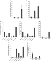
Colocalization of nanoparticles with rabankyrin-5, EEA-1, caveolin-1, clathrin, rab11, and Lamp-1. hCMEC/D3 endothelial cells were incubated with nanoparticles for 30 minutes at 4 °C. Subsequently, cells were incubated for (a–c) 30 minutes, (d,e) 60 minutes, (f) 90 minutes, and (g) 18 hours at 37 °C to allow particle internalization. (a) NBs and PrPBs do not colocalize with rabankyrin-5. A significant fraction of PEIBs is located in rabankyrin-5-positive vesicles. (b) NBs do not show significant colocalization with the early endosomal marker EEA-1, while 33% of the PrPBs show colocalization with EEA-1. Fifty-five percent of the PEIBs colocalize with EEA-1. (c) NBs and PrPBs show a similar extent of colocalization with caveolin-1, around 20%, and clathrin, <5%. In contrast, PEIBs colocalize extensively with clathrin. (d) The profile of colocalization of nanoparticles with the markers caveolin and clathrin at 60 minutes is comparable to that at earlier time points. (e) NBs and PrPBs lack rab11-colocalization. Around 30% of PEIBs reach a rab11-positive compartment after 60 minutes of internalization. (f) NBs are not identified within caveolin- or clathrin-positive compartments. PrPBs show an increase in colocalization with caveolin and clathrin in time. PEIBs continually colocalize with clathrin. (g) After 18 hours of incubation of hCMEC/D3 cells with nanoparticles, NBs and PEIBs sporadically colocalize with Lamp-1, whereas PrPBs are not detected in Lamp-1-positives vesicles. Cav, caveolin; clath, clathrin; EEA-1, early endosomal antigen-1; NB, noncoated bead; PEIB, polyethyleneimine-coated bead; PrPB, prion-coated bead.
Figure 4
Figure 4
NBs, PrPBs, and PEIBs localize in morphologically distinct intracellular vesicles. hCMEC/D3 cells were incubated with nanoparticles for 1 hour and 30 minutes. (a,b) NBs were found in vacuoles resembling multivesicular bodies (MVBs) and in multilamellar bodies. (c) PrPBs were primarily localized within vesicular structures. Note the presence of clathrin-coated buds (arrows) and the formation of clathrin latices around the prion coat (arrowheads). (d) PEIBs resided in large endosomes. Clathrin latices were found around the PEI coat (arrowheads). Bar = 500 nm. NB, noncoated bead; PEIB, polyethyleneimine-coated bead; PrPB, prion-coated bead.
Figure 5
Figure 5
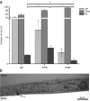
Transcytosis of nanoparticles across a monolayer of endothelial cells. (a) hCMEC/D3 cells were cultured on Transwell filters. Nanoparticles were added to the apical compartment and incubated for 18 hours. The total amount of iron was measured in the apical and basal compartment, and in the filter membrane with the cells. Transcytosis is presented as amount of iron in the apical compartment divided by the total amount of iron in the three compartments, and expressed as percentage (mean ± SEM, n = 3). Asterisks indicate significant differences (P < 0.05) in transcytosis of NBs versus PrPBs, PrPBs versus PEIBs, and NBs versus PEIBs, as evaluated with the Student's t-test. (b) Electron micrograph of a prion-coated particle (arrow) exocytosed at the abluminal side of a monolayer of hCMEC/D3 cells. AP, apical; BL, basal; NB, noncoated bead; PEIB, polyethyleneimine-coated bead; PrPB, prion-coated bead.

Similar articles

Cited by

References

    1. Wolf AA, Jobling MG, Wimer-Mackin S, Ferguson-Maltzman M, Madara JL, Holmes RK, et al. Ganglioside structure dictates signal transduction by cholera toxin and association with caveolae-like membrane domains in polarized epithelia. J Cell Biol. 1998;141:917–927. - PMC - PubMed
    1. Tuma PL., and, Hubbard AL. Transcytosis: crossing cellular barriers. Physiol Rev. 2003;83:871–932. - PubMed
    1. Muro S, Koval M., and, Muzykantov V. Endothelial endocytic pathways: gates for vascular drug delivery. Curr Vasc Pharmacol. 2004;2:281–299. - PubMed
    1. Norkin LC. Caveolae in the uptake and targeting of infectious agents and secreted toxins. Adv Drug Deliv Rev. 2001;49:301–315. - PubMed
    1. Predescu SA, Predescu DN., and, Malik AB. Molecular determinants of endothelial transcytosis and their role in endothelial permeability. Am J Physiol Lung Cell Mol Physiol. 2007;293:L823–L842. - PubMed