. 2010 Nov 2:1:109.
doi: 10.1038/ncomms1108.

TRPM7 is essential for Mg(2+) homeostasis in mammals

Affiliations
Free PMC article

TRPM7 is essential for Mg(2+) homeostasis in mammals

Lillia V Ryazanova et al. Nat Commun. .
Free PMC article

Abstract

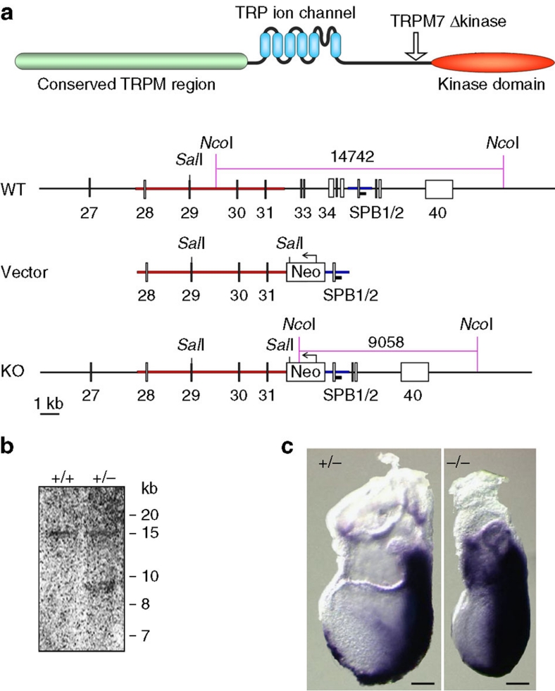
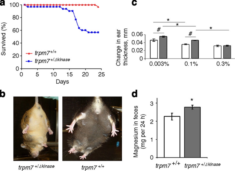
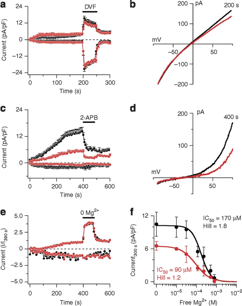
Mg(2+) is the second-most abundant cation in animal cells and is an essential cofactor in numerous enzymatic reactions. The molecular mechanisms controlling Mg(2+) balance in the organism are not well understood. In this study, we report identification of TRPM7, a bifunctional protein containing a protein kinase fused to an ion channel, as a key regulator of whole body Mg(2+) homeostasis in mammals. We generated TRPM7-deficient mice with the deletion of the kinase domain. Homozygous TRPM7(Δkinase) mice demonstrated early embryonic lethality, whereas heterozygous mice were viable, but developed signs of hypomagnesaemia and revealed a defect in intestinal Mg(2+) absorption. Cells derived from heterozygous TRPM7(Δkinase) mice demonstrated reduced TRPM7 currents that had increased sensitivity to the inhibition by Mg(2+). Embryonic stem cells lacking TRPM7 kinase domain displayed a proliferation arrest phenotype that can be rescued by Mg(2+) supplementation. Our results demonstrate that TRPM7 is essential for the control of cellular and whole body Mg(2+) homeostasis.


Figures

Figure 1
Figure 1. Generation of TRPM7-deficient mice.
(a) A schematic representation of TRPM7 protein. Arrow indicates the position of truncation in TRPM7Δkinase mice. Also shown are the structures of the murine TRPM7 locus, targeting vector and resulting homologous recombination locus. (b) Southern blot analysis of genomic DNA from wild-type (+/+) or TRPM7+/Δkinase (+/−) mice after digestion with NcoI. (c) TRPM7+/Δkinase (+/−) and TRPM7Δkinase/Δkinase (−/−) embryos at 7.5 days post coitum. Staining with Brachyury antisense mRNA marks the primitive streak, where mesoderm formation occurs, and demonstrates that embryos are at the advanced primitive streak stage. Scale bar, 100 μm.
Figure 2
Figure 2. Analysis of hypomagnesaemic phenotype in TRPM7+/Δkinase mice.
(a) Survival of wild-type (TRPM7+/+) mice (n=30) or TRPM7+/Δkinase mice (n=30) on Mg2+-deficient diet. (b) Clasping test for the wild-type and TRPM7+/Δkinase mice subjected to Mg2+-free diet for 12 days. (c) The effect of TRPM7 deficiency and Mg2+ content in the diet on the oxazolone sensitivity reaction. The delayed sensitivity response is measured as a change in the ear thickness, mm. Before ear application of oxazolone, wild-type mice and TRPM7+/Δkinase mice were fed for 7 days severely Mg2+ deficient, control and supplemental Mg2+ diet with 0.003, 0.1 and 0.3% Mg2+, respectively. The swelling response was measured 24 h after the challenge using an engineer's micrometre. Calculations of the swelling response were carried out comparing with the respective control groups not exposed to oxazolone. All mice were 4 months old. Open and shaded bars correspond to wild-type mice and TRPM7+/Δkinase mice respectively. Values are means ± s.e.m. Two-way analysis of variance test was used for the calculations (n=7–10 mice per group); P<0.05. #Significantly different from wild-type mice fed a respective diet. *Significantly different from respective wild-type or TRPM7+/Δkinase mice fed a control 0.1% Mg diet. (d) Analysis of Mg2+ content in feces of the wild type mice (n=6) and TRPM7+/Δkinase mice (n=6). Mice were maintained on normal diet (0.1% Mg2+). Values are means ± s.e.m. Statistical analysis was performed by Student's t-test. *Significantly different (P<0.05).
Figure 3
Figure 3. TRPM7 (MagNuM) currents in mast cells derived from TRPM7-deficient mice.
(a) Average normalized time course of MagNuM current development in mouse mast cells isolated from wild-type (black circles, n=15) and TRPM7+/Δkinase mice (red circles, n=11). Currents were elicited by a ramp protocol from −100 to +100 mV over 50 ms acquired at 0.5 Hz. Inward current amplitudes were extracted at −80 mV, outward currents at +80 mV and plotted versus time of the experiment. Data were normalized to cell size as pA/pF. Error bars indicate s.e.m. The composition of extracellular, intracellular and application solutions are described in Methods section. (b) Average current–voltage (I/V) curves extracted from a representative wild-type cell (black) and TRPM7+/Δkinase (red) extracted at 200 s. (c) Average normalized time course of MagNuM current development in mouse mast cells isolated from wild-type (black circles, n=10) and TRPM7+/Δkinase mice (red circles, n=12). Data were acquired and analysed as in a. At the time indicated by the black bar, cells were superfused with external solution supplemented with 50 μM 2-APB. (d) Current–voltage (I/V) curves extracted from a representative wild-type cell (black) and TRPM7+/Δkinase (red). (e) Average normalized time course of MagNuM current development in mouse mast cells isolated from wild-type (black circles, n=14) and TRPM7+/Δkinase mice (red circles, n=9). Data were acquired as in a and normalized as I/I390 s. Experimental solutions as in a, except for the application solution that contained (in mM) 150 NaCl, 2.8 KCl, 3 CaCl2, 10 HEPES–NaOH. Extracellular solution was nominally Mg2+ free, and applied for the time indicated by the black bar. Internal solution contained 10 mM Cs-BAPTA instead of Cs-EGTA. (f) Dose–response curve of MagNuM currents to intracellular Mg2+ measured in mouse mast cells isolated from wild-type (black circles, n=5–11) or TRPM7+/Δkinase mice (red circles, n=5–10). Error bars indicate s.e.m. Current amplitudes assessed at +80 mV and extracted at 300 s into the experiment. Intracellular Mg was (in μM) 0 (nominally free), 40 (31 free), 150 (116 free), 300 (233 free), 650 (507 free), 1,000 (781 free), and was calculated using WebMAXC.
Figure 4
Figure 4. TRPM7 (MagNuM) currents in ES cells derived from TRPM7 deficient mice.
(a) Average normalized time course of TRPM7 current development in mouse ES cells isolated from wild-type (black circles, n=13), TRPM7+/Δkinase (red circles, n=12) and TRPM7Δkinase/Δkinase mice (blue circles, n=13). Data were acquired and analysed as in Figure 3a. Error bars indicate s.e.m. The intracellular solution was divalent-free and contained (in mM) 140 Cs-glutamate, 8 NaCl, 10 Cs-EGTA, 5 Na-EDTA (pH 7.2, 300 mOsm). The extracellular solution contained (in mM) 140 NaCl, 1 CaCl2, 2.8 KCl, 2 MgCl2, 10 HEPES–NaOH, 11 glucose (pH 7.2, 300 mOsm). The application solution contained (in mM) 150 mM NaCl, 2.8 KCl, 10 HEPES–NaOH, 0.5 Na-EDTA. (b) Average current–voltage (I/V) curves from a representative wild-type cell (black), TRPM7+/Δkinase (red) and TRPM7Δkinase/Δkinase (blue) extracted at 200 s. (c) Average normalized time course of MagNuM current development in mouse ES cells isolated from wild-type (black circles, n=8), TRPM7+/Δkinase mice (red circles, n=10) and TRPM7Δkinase/Δkinase mice (blue circles, n=10). Data were acquired and analysed as in a. At the time indicated by the black bar, cells were superfused with external solution supplemented with 50 μM 2-APB. (d) Current–voltage (I/V) curves extracted from a representative wild-type cell (black), TRPM7+/Δkinase (red) and TRPM7Δkinase/Δkinase (blue).
Figure 5
Figure 5. Mg2+ supplementation rescues the growth arrest phenotype of TRPM7 deficient ES cells.
Equal amounts (2.2 × 10⁵ per well) of wild-type, TRPM7+/Δkinase and TRPM7Δkinase/Δkinase ES cells were plated in six-well plates in 2 ml of the control ES-DMEM and ES-DMEM supplemented with 10 mM MgCl2. (a) Phase-contrast images were taken after 72 h of growth. Scale bar, 200 μm. (b) Growth curves. Number of ES cells were quantified at different time points. Error bars indicate s.e.m., n=3.

References

    1. Schmitz C. et al.. Regulation of vertebrate cellular Mg2+ homeostasis by TRPM7. Cell 114, 191–200 (2003). - PubMed
    1. Runnels L. W., Yue L. & Clapham D. E. TRP-PLIK, a bifunctional protein with kinase and ion channel activities. Science 291, 1043–1047 (2001). - PubMed
    1. Nadler M. J. et al.. LTRPC7 is a Mg.ATP-regulated divalent cation channel required for cell viability. Nature 411, 590–595 (2001). - PubMed
    1. Schlingmann K. P. et al.. Hypomagnesemia with secondary hypocalcemia is caused by mutations in TRPM6, a new member of the TRPM gene family. Nat. Genet. 31, 166–170 (2002). - PubMed
    1. Walder R. Y. et al.. Mutation of TRPM6 causes familial hypomagnesemia with secondary hypocalcemia. Nat. Genet. 31, 171–174 (2002). - PubMed