Review
. 2010 Dec;11(12):812-22.
doi: 10.1038/nrn2924. Epub 2010 Nov 3.

The diverse roles of ribbon synapses in sensory neurotransmission

Affiliations
Review

The diverse roles of ribbon synapses in sensory neurotransmission

Gary Matthews et al. Nat Rev Neurosci. 2010 Dec.

Abstract

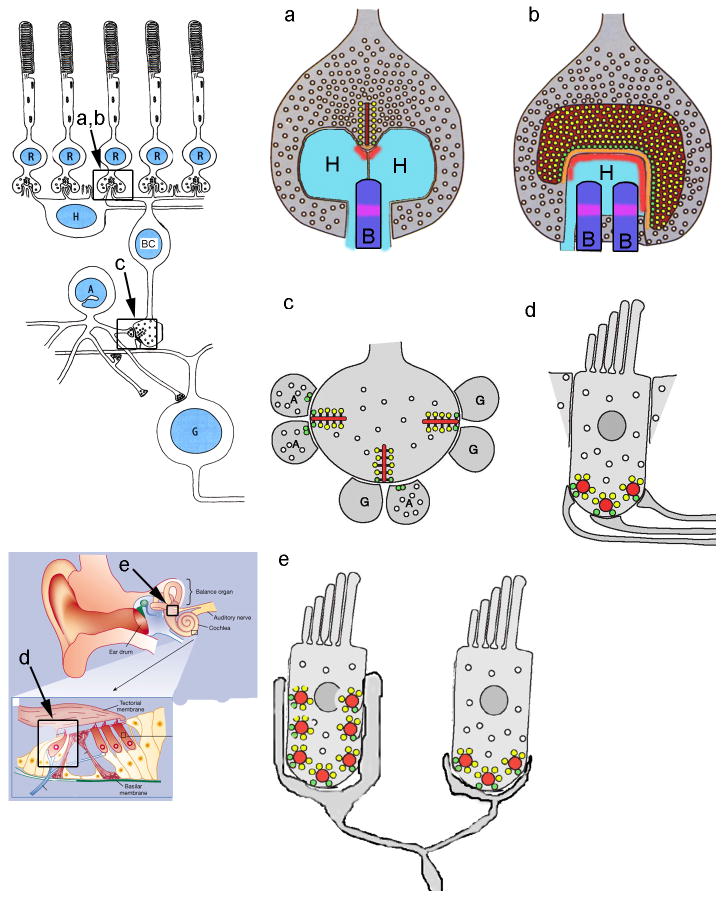
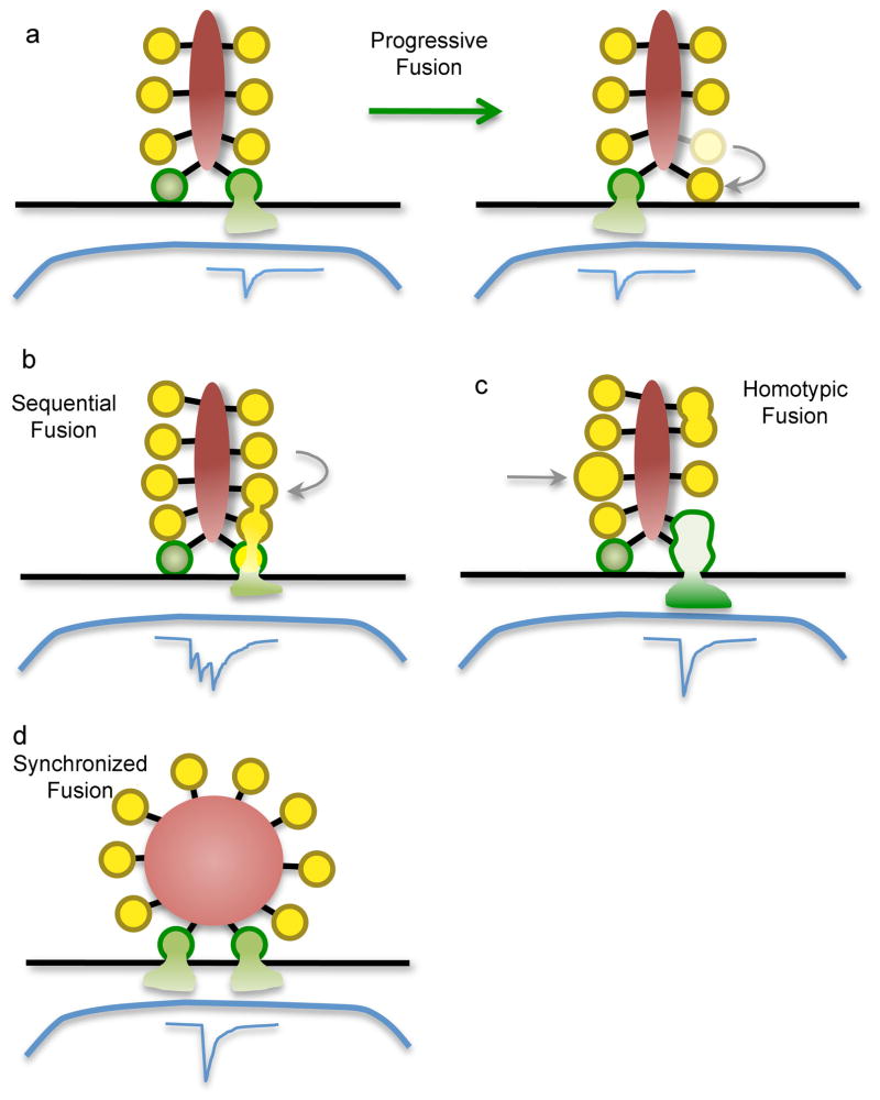
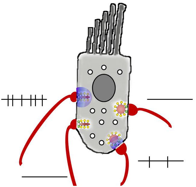
Sensory synapses of the visual and auditory systems must faithfully encode a wide dynamic range of graded signals, and must be capable of sustained transmitter release over long periods of time. Functionally and morphologically, these sensory synapses are unique: their active zones are specialized in several ways for sustained, rapid vesicle exocytosis, but their most striking feature is an organelle called the synaptic ribbon, which is a proteinaceous structure that extends into the cytoplasm at the active zone and tethers a large pool of releasable vesicles. But precisely how does the ribbon function to support tonic release at these synapses? Recent genetic and biophysical advances have begun to open the 'black box' of the synaptic ribbon with some surprising findings and promise to resolve its function in vision and hearing.


Figures

Figure 1
Figure 1. Diversity of ribbon morphology and postsynaptic architecture in different cell types
a, b: Schematic diagram of transverse (a) and en-face (b) views of a mammalian photoreceptor ribbon synapse, whose context in the retina is illustrated in the diagram to the left. c: Synaptic arrangement at bipolar cell ribbons. The location of this synapse in retinal circuitry is illustrated in the diagram to the left. d: Synaptic arrangement at ribbons of cochlear inner hair cells. e: Vestibular afferents receive synaptic inputs from multiple ribbons of multiple hair cells. The locations of cochlear inner hair cells and the vestibular apparatus are shown schematically in the diagram to the left. Dark red areas, synaptic ribbons; bright red areas, AMPA receptors; orange areas, arciform density; yellow circles, vesicles attached to ribbon; green circles, docked vesicles; light blue areas, horizontal cell dendrites (H); dark blue areas, ON bipolar cell dendrites (B); magenta areas, mGluR6 receptors; G: ganglion cell dendrite; A: amacrine cell process.
Figure 2
Figure 2. Modes of synaptic vesicle fusion at ribbon synapses
Green vesicles represent vesicles that are docked at the plasma membrane (black line) at release sites. Blue traces represent the postsynaptic current. a: Progressive fusion, in which primed vesicles docked at different release sites fuse progressively during maintained depolarization, each producing an independent unitary postsynaptic current. Vesicles in higher rows move along the ribbon to replenish empty release sites. b,c: Two forms of compound fusion. In sequential fusion (b), vesicles fuse in sequence up the face of the ribbon, starting with the vesicles docked at the plasma membrane. This produces a burst of quantal postsynaptic currents in rapid sequence. Vesicles attached to the ribbon might fuse with each other (homotypic fusion; c) prior to undergoing exocytosis. The resulting larger bolus of glutamate produces a larger postsynaptic current. d: Synchronized fusion of multiple docked vesicles, which results in a multiquantal postsynaptic current.
Figure 3
Figure 3. Variable ribbon function within one cochlear inner hair cell
‘Low threshold’ ribbons may have greater numbers of calcium channels, release transmitter more frequently at a given membrane potential, and so trigger more afferent action potentials. ‘High threshold’ ribbons may have fewer calcium channels and therefore require greater depolarization to cause equivalent levels of afferent activity. Alternatively, variations in vesicular fusion (e.g., different sensitivities to calcium or coordination of multivesicular release) could produce similar outcomes. Overall probability of transmission is relatively low, so not every synapse will be active at a given point in time. The purple cloud indicates calcium influx; lines indicate action potentials in afferent fibres.
Box Figure
Box Figure

References

    1. Dick O, tom Dieck S, Altrock WD, Ammermuller J, Weiler R, Garner CC, et al. The presynaptic active zone protein bassoon is essential for photoreceptor ribbon synapse formation in the retina. Neuron. 2003;37:775–786. - PubMed
    1. Heidelberger R, Thoreson WB, Witkovsky P. Synaptic transmission at retinal ribbon synapses. Prog Retina Eye Res. 2005;24:682–720. - PMC - PubMed
    1. Sterling P, Matthews G. Structure and function of ribbon synapses. Trends Neurosci. 2005;28:20–29. - PubMed
    1. DeVries SH, Li W, Saszik S. Parallel processing in two transmitter microenvironments at the cone photoreceptor synapse. Neuron. 2006;50:735–748. - PubMed
    1. von Gersdorff H, Vardi E, Matthews G, Sterling P. Evidence that vesicles on the synaptic ribbon of retinal bipolar neurons can be rapidly released. Neuron. 1996;16:1221–1227. - PubMed

Publication types

MeSH terms

Substances