

Transport of L-carnitine in human corneal and conjunctival epithelial cells
- PMID: 21045919
- PMCID: PMC2956661
Transport of L-carnitine in human corneal and conjunctival epithelial cells
Abstract
Purpose: Previously we demonstrated expression and localization of carnitine/organic cation transporters, OCTN1 and OCTN2, in human corneal and conjunctival epithelia. The present study aimed to examine the characteristics of L-carnitine transporters in cultured human limbal corneal (HCLE) and conjunctival epithelial (HCjE) cells.
Methods: Time-course, Na(+)-dependence, kinetics, energy- and pH- dependence of L-carnitine transport were investigated by monitoring L-[(3)H]carnitine uptake into HCLE and HCjE cells. To determine the specificity of action, competition and inhibition studies were performed.
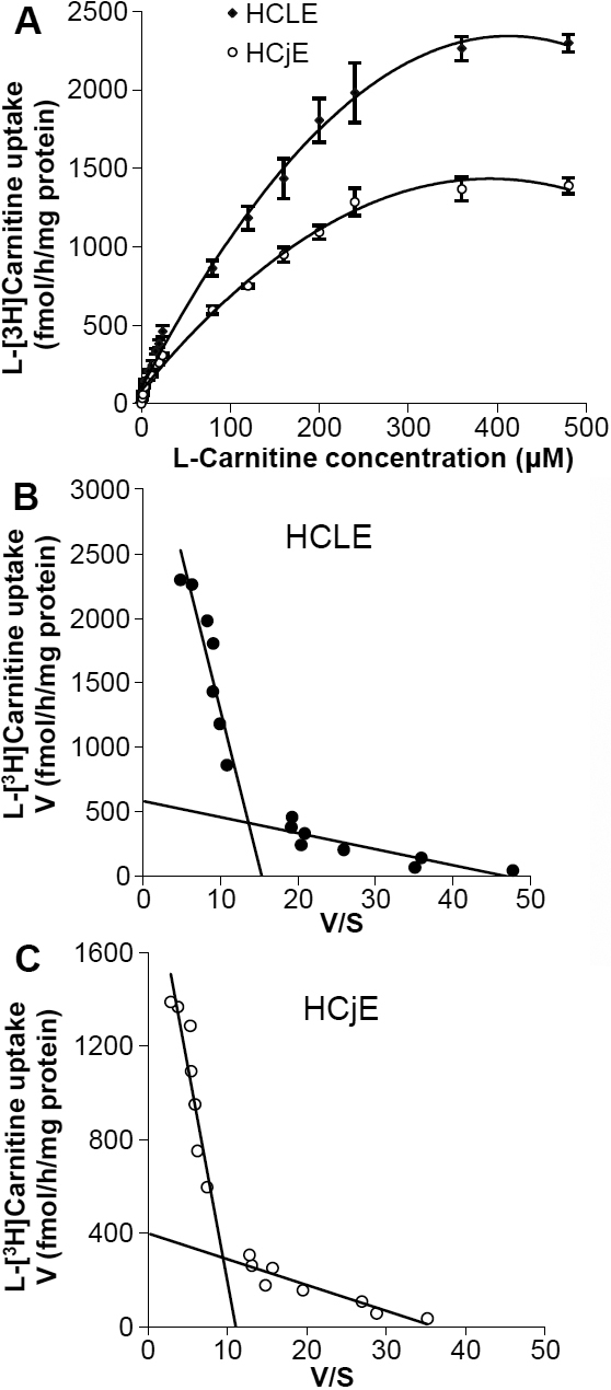
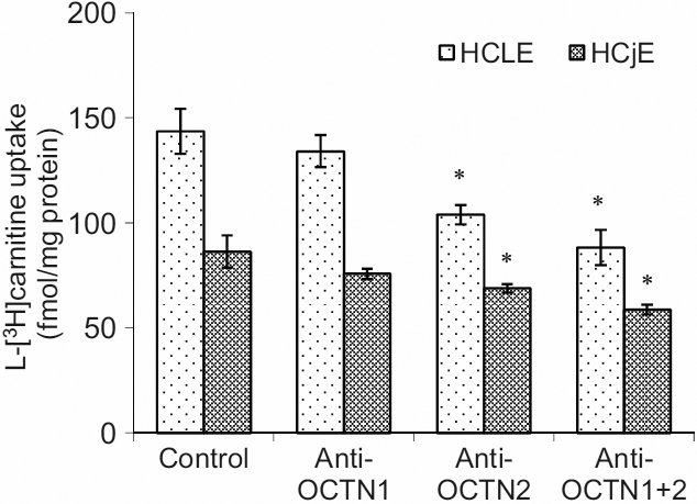
Results: The uptake of L-carnitine into HCLE and HCjE cells was saturable and time-dependent. An Eadie-Hofstee plot showed two distinct components: a high- and a low- affinity carnitine transport system in HCLE and/or HCjE cells. L-carnitine transport was significantly inhibited by the metabolic inhibitors (sodium azide, dinitrophenol, iodoacetic acid). The L-carnitine analogs (D-carnitine, acetyl-L-carnitine and γ-butyrobetaine), tetraethylammonium (TEA), 2-amino-2-norbornane carboxylic acid (BCH), strongly inhibited uptake of L-[(3)H]carnitine. Uptake of L-[(3)H]carnitine also required the presence of Na(+) in the external medium and the uptake activity was maximal at pH 5.5. The anti-OCTN2 antibody blocked L-carnitine uptake in both HCLE and HCjE cells whereas the anti-OCTN1 antibody did not significantly block L-carnitine uptake.
Conclusions: L-carnitine is transported into HCLE and HCjE cells by an active carrier mediated transport system that is time-, Na(+)-, energy- and pH- dependent. The carnitine/organic cation transporter OCTN2 appears to play a dominant role in this process.
Figures


Similar articles
-
Expression and localization of carnitine/organic cation transporter OCTN1 and OCTN2 in ocular epithelium.Invest Ophthalmol Vis Sci. 2008 Nov;49(11):4844-9. doi: 10.1167/iovs.07-1528. Epub 2008 Jul 18. Invest Ophthalmol Vis Sci. 2008. PMID: 18641280
-
Characteristics of L-carnitine transport in cultured human hepatoma HLF cells.J Pharm Pharmacol. 1999 Aug;51(8):935-40. doi: 10.1211/0022357991773195. J Pharm Pharmacol. 1999. PMID: 10504033
-
Molecular and functional identification of sodium ion-dependent, high affinity human carnitine transporter OCTN2.J Biol Chem. 1998 Aug 7;273(32):20378-82. doi: 10.1074/jbc.273.32.20378. J Biol Chem. 1998. PMID: 9685390
-
Carnitine transport: pathophysiology and metabolism of known molecular defects.J Inherit Metab Dis. 2003;26(2-3):147-69. doi: 10.1023/a:1024481016187. J Inherit Metab Dis. 2003. PMID: 12889657 Review.
-
Characterization of organic cation/carnitine transporter family in human sperm.Biochem Biophys Res Commun. 2003 Jun 20;306(1):121-8. doi: 10.1016/s0006-291x(03)00930-6. Biochem Biophys Res Commun. 2003. PMID: 12788076 Review.
Cited by
-
Decrease in hyperosmotic stress-induced corneal epithelial cell apoptosis by L-carnitine.Mol Vis. 2013 Sep 19;19:1945-56. eCollection 2013. Mol Vis. 2013. PMID: 24068862 Free PMC article.
-
Tear Metabolomics in Dry Eye Disease: A Review.Int J Mol Sci. 2019 Aug 1;20(15):3755. doi: 10.3390/ijms20153755. Int J Mol Sci. 2019. PMID: 31374809 Free PMC article. Review.
-
Distribution, molecular structure and functional analysis of carnitine transporter (SLC22A5) in canine lens epithelial cells.Exp Anim. 2014;63(4):467-73. doi: 10.1538/expanim.63.467. Epub 2014 Jul 22. Exp Anim. 2014. PMID: 25048262 Free PMC article.
-
Transporter-Targeted Nano-Sized Vehicles for Enhanced and Site-Specific Drug Delivery.Cancers (Basel). 2020 Oct 1;12(10):2837. doi: 10.3390/cancers12102837. Cancers (Basel). 2020. PMID: 33019627 Free PMC article. Review.
-
Categorization of Marketed Artificial Tear Formulations Based on Their Ingredients: A Rational Approach for Their Use.J Clin Med. 2021 Mar 21;10(6):1289. doi: 10.3390/jcm10061289. J Clin Med. 2021. PMID: 33800965 Free PMC article.
References
-
- Pessotto P, Liberati R, Petrella O, Romanelli L, Calvani M, Peluso G. In experimental diabetes the decrease in the eye of lens carnitine levels is an early important and selective event. Exp Eye Res. 1997;64:195–201. - PubMed
-
- Corrales RM, Luo L, Chang EY, Pflugfelder SC. Effects of osmoprotectants on hyperosmolar stress in cultured human corneal epithelial cells. Cornea. 2008;27:574–9. - PubMed
-
- Garrett Q, Min-Hsuan Shih S, Simmons PA, Vehige J, Willcox M. Carnitine and the Potential Osmoprotectants Protect Corneal Epithelial Cells from Hyperosmolar Solution Induced Damage. ARVO Annual Meeting; 2010 May 2–6; Fort Lauderdale (FL).
-
- Simmons P, Chang-Lin J, Chung Q, Vehige J, Carlisle-Wilcox C, Welty D. Selection of compatible solutes for inclusion in a lubricant eye drop. TFOS meeting 2007 September, 2007; Sicily, Italy.
-
- Kerner J, Hoppel C. Genetic disorders of carnitine metabolism and their nutritional management. Annu Rev Nutr. 1998;18:179–206. - PubMed
Publication types
MeSH terms
Substances
LinkOut - more resources
Full Text Sources