Review
. 2010:30:119-30.

Total ankle replacement: why, when and how?

Affiliations
Review

Total ankle replacement: why, when and how?

Davide Edoardo Bonasia et al. Iowa Orthop J. 2010.

Abstract

Total ankle replacement (TAR) was first attempted in the 1970s, but poor results led to its being considered inferior to ankle fusion until the late 1980s and early 1990s. By that time, newer designs which more closely replicated the natural anatomy of the ankle, showed improved clinical outcomes. Currently, even though controversy still exists about the effectiveness of TAR compared to ankle fusion, TAR has shown promising mid-term results and should no longer be considered an experimental procedure. Factors related to improved TAR outcomes include: 1) better patient selection, 2) more precise knowledge and replication of ankle biomechanics, 3) the introduction of less-constrained designs with reduced bone resection and no need for cementation, and 4) greater awareness of soft-tissue balance and component alignment. When TAR is performed, a thorough knowledge of ankle anatomy, pathologic anatomy and biomechanics is needed along with a careful pre-operative plan. These are fundamental in obtaining durable and predictable outcomes. The aim of this paper is to outline these aspects through a literature review.


Figures
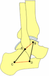
Figure 1
Figure 1
Two-dimensional four-bar linkage model of the ankle joint to describe dorsi/plantar flexion in unloaded conditions. The talus/ calcaneus complex and tibia/fibula complex rotate about each other on approximately inextensible line segments, represented by the calcaneofibular (AC) and tibiocalcaneal (BD) ligaments. The other two bars in this four-bar linkage model are formed by: 1) the bony segment connecting the proximal insertions of calcaneofibular and tibiocalcaneal ligaments (AB, red dashed line); and 2) the bony segment connecting the distal insertions of calcaneofibular and tibiocalcaneal ligaments (CD, red dashed line). Redrawn from Leardini etal. J Biomech. 1999 Jun;32(6):585-91.
Figure 2
Figure 2
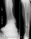
Left post-traumatic ankle arthritis. A) Cobey-Saltzman view. B) Antero-posterior radiograph of the left leg showing a varus deformity, with center of rotation of angulation (CORA) located at the distal third of the tibia.
Figure 3
Figure 3
Pre-operative measurement. A) The lateral distal tibial angle (LDTA) is formed by the distal tibial articular surface and the anatomical axis of the tibia (normal values 89° ± 3°). B) The anterior distal tibial angle (ADTA) is formed by the mechanical axis of the tibia and the joint orientation line of the ankle in the sagittal plane (normal values 80° ± 3°). C) The tibial-talar angle (a) is defined by the tibial and talar articular surfaces in the ankle joint; if it measures > 10°, the joint is defined as incongruent.
Figure 4
Figure 4
The center of rotation of angulation (CORA) is the intersection of the mid-diaphyseal line and the line starting from the middle of the joint and perpendicular to the abnormal ADTA or LDTA (LDTA in this figure). The CORA can be located proximally at the tibia (A) or at the joint line level (B).
Figure 5
Figure 5
Post-traumatic left ankle arthritis in a varus congruent joint (same patient as Figure 2). A) Careful debridement of the osteophytes, synovial tissue and excessive capsule. B) Tibial guide positioning under fluoroscopy in order to obtain a neutralizing tibial cut (greater bone removal on the lateral side) to restore alignment. Even though the CORA is diaphyseal (see Figure 2), malalignment is minimal (6°) and can be addressed with the TAR, with no need for osteotomy
Figure 6
Figure 6
Post-traumatic left ankle arthritis in a varus congruent joint (same patient as Figure 2 and 5). A) After tibial and talar cuts, soft tissue balance is checked with a laminar spreader. B) After trial component positioning, the range of motion is checked. If there is insufficient dorsiflexion (not due to component malpositioning) percutaneous Achilles tendon lengthening is performed. C) Final components of Salto Talaris TAR implanted.
Figure 7
Figure 7
Post-operative fluoroscopy. A) Lateral view, B) Antero-posterior view, showing a valgus correction of 7° on the coronal plane, in order to correct the 6° of diaphyseal varus deformity (see Figure 2).

Similar articles

Cited by

References

    1. Vickerstaff JA, Miles AW, Cunningham JL. A brief history of total ankle replacement and a review of the current status. Med Eng Phys. 2007;29(10):1056–64. Dec. - PubMed
    1. Chou LB, Coughlin MT, Hansen S, Jr, Haskell A, Lundeen G, Saltzman CL, Mann RA. Osteoarthritis of the ankle: the role of arthroplasty. J Am Acad Orthop Surg. 2008;16(5):249–59. May. - PubMed
    1. Inman VT. The joints of the ankle. Baltimore: Williams&Wilkins; 1979.
    1. Isman RE, Inman VT. Anthropometric studies of the human foot and ankle. Bull Pros Res. 1969;10:97–129.
    1. Bottlang M, Marsh JL, Brown TD. Articulated external fixation of the ankle: minimizing motion resistance by accurate axis alignment. J Biomech. 1999;32:63–70. - PubMed

LinkOut - more resources